
10 อันดับเว็บพนันออนไลน์ไม่ผ่านเอเย่นต์
พบรีวิวผู้ให้บริการคาสิโนที่ดีที่สุด รวม 10 อันดับเว็บพนันออนไลน์ไม่ผ่านเอเย่นต์ชั้นนำที่เชื่อถือได้เท่านั้น พร้อมข้อมูลจริงที่เปิดโอกาสให้คุณเป็นส่วนหนึ่งของเกมการเดิมพันที่สร้างทั้งความบันเทิงและกำไรอย่างเป็นกอบเป็นกำ โดยไกด์บนหน้านี้จะช่วยให้คุณเลือกเวปตรงที่ได้คุณภาพ พร้อมรับประสบการณ์การเดิมพันกับเจ้ามือที่ดีและตรงกับสิ่งที่คุณมองหาที่สุด
เว็บตรงไม่ผ่านเอเย่นต์ 10 แบรนด์กับลิสต์โบนัสคุ้มๆ
1 |  M88 ⭐⭐⭐⭐⭐ | M88 โบนัสต้อนรับกีฬา 125% สูงถึง 3,500 บาท ✅ พนันสะดวก วางบิลง่ายใน 5 นาที ✅ รับเครดิตฝากไว แทงบอลสดทันใจ ✅ เกมจ่ายคืนสูง มีให้เล่นเฉพาะที่ M88 | |
2 |  WE88 ⭐⭐⭐⭐⭐ | we88 โบนัสล็อต 100% สูงถึง 5,000 บาท ✅ ทัวร์นาเมนต์ Daily Wins รางวัลหลักล้าน ✅ WeSpin ให้โอกาสลุ้นเงินสดง่ายๆ รายวัน ✅ เวปตรงที่เป็นแหล่งเกม P2P ที่ดีที่สุดในไทย | |
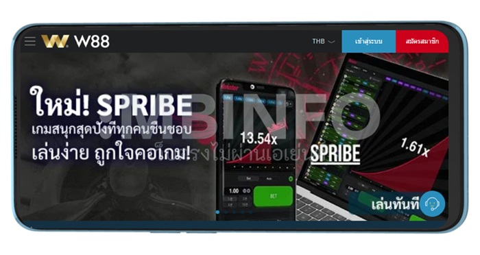
3 |  W88 ⭐⭐⭐⭐⭐ | W88 โปรโมชั่น ต้อนรับคอหวย 100% ✅ เปิดให้เล่นเกมใหม่เล่นง่ายตลอดปี ✅ หนึ่งในเว็บตรงที่เปิดให้เล่นโป๊กเกอร์ ✅ มีโบนัสให้เลือกหลายรายการที่สุด | |
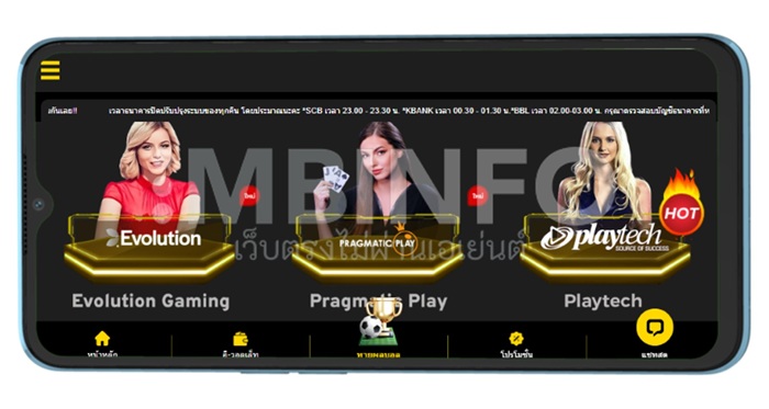
4 |  12PLAY ⭐⭐⭐⭐⭐ | 12play เงินคืนเกมสล็อตไม่จำกัด ทุกวัน 1.2% ✅ จ่ายเงินไว ปลอดภัย และไม่ยุ่งยาก ✅ เปิดตัวค่ายสล็อตใหม่ๆ โบนัสเพียบ ✅ โอกาสร่วมทัวร์นาเมนต์ลุ้นรางวัลใหญ่ | |
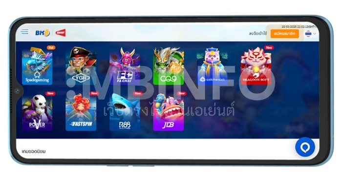
5 |  BK8 ⭐⭐⭐⭐ | BK8 ต้อนรับสมาชิกใหม่ ฝาก 500 ฟรี 500 บาท ✅ คาสิโนคริปโตดีที่สุดในไทย ได้มาตรฐาน ✅ ถอนเงินง่ายและรวดเร็ว รับภายใน 15 นาที ✅ แทงหวย 2-3 ตัว อัตราจ่ายออกสูงสุดในไทย | |
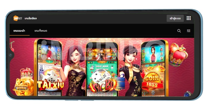
6 |  188BET ⭐⭐⭐⭐ | 188bet โบนัสบอลสเต็ปแจกหนัก จัดเต็มที่สุด ✅ ให้โบนัสทอนคอมรายสัปดาห์ รับง่ายได้จริง ✅ แจกโปรหลายรายการ ผู้เล่นรับได้ทั่วถึง ✅ แหล่งรวมเกมสล็อตแจ็คพ็อตหาเล่นง่าย | |
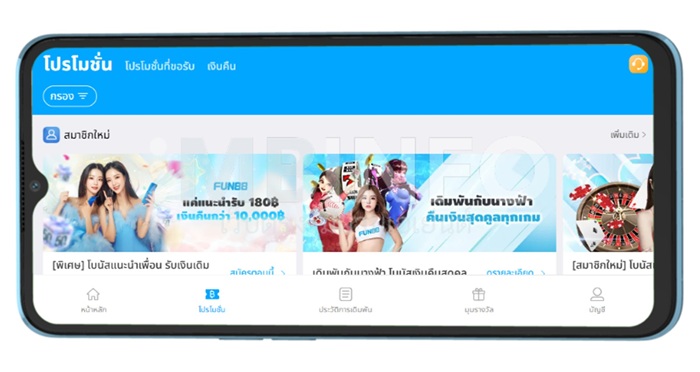
7 |  FUN88 ⭐⭐⭐⭐ | Fun88 สมาชิกใหม่ โบนัสแรกเข้า 100% คาสิโน ✅ กิจกรรมน่าสนใจ “มุมรางวัล” สุดคุ้มค่า ✅ เนทีฟแอป FUN88 ประสบการณ์ดีที่สุด ✅ แหล่งเวปตรงรวมเกมฮอต เกมจ่ายสูง | |
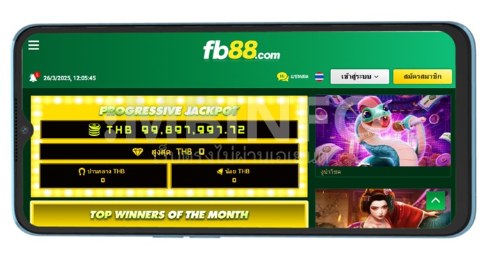
8 |  FB88 ⭐⭐⭐ | FB88 โบนัสต้อนรับ 150% ในสล็อต ✅ ฝากเงินรับเครดิตใน 2 นาที จ่ายไวใน 5 นาที ✅ รับพนันด้วยคริปโต ทันสมัยและปลอดภัย ✅ แลกรับรางวัลพรีเมี่ยมที่คลับรางวัล FB88 | |
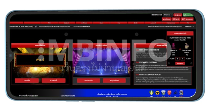
9 |  12BET ⭐⭐⭐ | 12bet เวปตรงแจก 120% โบนัสยินดีต้อนรับ ✅ 12bet 2.0 ประสบการณ์ใหม่ โหลดไวขึ้น ✅ โปรแกรมพิเศษ 12Rewards เฉพาะสมาชิก ✅ เว็บพนันตรงบนมือถือ โหลดไว เล่นง่าย | |
10 |  JBO ⭐⭐⭐ | JBO สมาชิกใหม่รับ 3,000 บาท + 2,000 ฟรีสปิน ✅ พัฒนาแอปฯ พนันบนมือถือทุกเกมดัง ✅ ลุ้นมินิเกม ไทยไฮโล, น้ำเต้าปูปลา, บาคาร่า ✅ ไฮไลท์ AG คาสิโนสด ไลฟ์ดีลเลอร์สุดฮอต | |
11 | Alpha88 ⭐⭐⭐ | โปรโมชั่นต้อนรับ GPI คาสิโน โบนัส 100% สุดคุ้ม! ✅ กีฬา คาสิโน สล็อต ครบจบในเว็บเดียว ✅ ฝากถอนเร็วใน 1-5 นาที ปลอดภัยทุกขั้นตอน ✅ รองรับคริปโต เล่นง่ายจ่ายคล่อง | |
12 | Y88 ⭐⭐⭐ | ฝากเงินครบสองครั้งกับ Y88 รับโบนัสไปเลย ✅ ไม่ผ่านเอเย่นต์ ปลอดภัย มั่นใจทุกการเดิมพัน ✅ มีค่ายเกมสล็อตให้เลือกเล่นมากกว่า 30 ค่าย ✅ เกมสล็อตแตกง่าย คัดมาเฉพาะเกมที่มี RTP สูงและฟรีสปินบ่อย | |
13 | Nova88 ⭐⭐⭐ | 100% Kick-Off โบนัสต้อนรับจาก Nova88 ✅ เดิมพันกีฬากับ SABA Sports ครบทุกลีกทั่วโลก ✅ รองรับมือถือ ใช้งานได้ทุกระบบ ✅ มีเกมและกีฬาหลากหลายครบวงจร | |
14 | Live Casino House ⭐⭐⭐ | โปรต้อนรับคาสิโน 100% สูงถึง 4,000 บาท ✅ เกมสล็อตและคาสิโนสดครบทุกค่ายดัง ✅ รับเงินคืนรายวันและรายสัปดาห์ ไม่จำกัด ✅ โปรแนะนำเพื่อน รับเงินเพิ่มฟรี | |
15 | HUC99 ⭐⭐⭐ | HUC99 ชวนเพื่อนมาเล่น รางวัลสูงสุด 9999 บาท ✅ สมาชิกใหม่รับเครดิตฟรี 1299 บาท และฟรีสปินสล็อต ✅ ระบบฝาก-ถอนอัตโนมัติ เร็ว ปลอดภัย ไม่ต้องผ่านแอดมิน ✅ ดาวน์โหลดแอป HUC99 ใช้งานสะดวกทุกที่ทุกเวลา | |
16 | HappyLuke ⭐⭐⭐ | โบนัสต้อนรับ 100% คาสิโน สูงสุด 5,000 บาท ✅ โบนัสรายวัน รายสัปดาห์ รับสูงสุด 30% ✅ เควสรายวันและกิจกรรมพิเศษ ลุ้นโบนัสทุกวัน ✅ บริการลูกค้าตลอด 24 ชม. พร้อมช่วยเหลือทุกข้อสงสัย | |
17 | IB8 ⭐⭐⭐ | โบนัสต้อนรับสมาชิกทุกท่าน 666 บาท ✅ ดาวน์โหลดแอปวันนี้ รับโบนัสฟรี 50 บาท ✅ กิจกรรมพิเศษมากมาย เช่น หมุนนำโชค, เช็คอินรายวัน, อั่งเปา ✅ ระบบ VIP พร้อมสิทธิพิเศษเฉพาะระดับสมาชิก | |
18 | Kapook888 ⭐⭐⭐ | โปรวัดใจ Baccarat แทงถูกผิดติดต่อกัน 12 ครั้งรับทันที 88888 บาท ✅ เดิมพันกีฬาให้เลือกมากกว่า 25,000 รายการต่อเดือน ✅ ปลอดภัย มั่นใจด้วยใบรับรองจาก BMM, iTech Labs, GoDaddy ✅ รับเงินคืนทุกวัน 1% สำหรับกีฬา, E-sport, คาสิโน, สล็อต, ยิงปลา | |
19 | LuckyDays ⭐⭐⭐ | โบนัสเงินฝาก 100% และฟรีสปิน 33 ครั้ง ✅ โบนัสต้อนรับสูงสุด 6,300 บาท + 99 ฟรีสปิน สำหรับ 3 ครั้งแรก ✅ ความปลอดภัยสูง มีใบอนุญาตจาก Curacao พร้อมมาตรฐาน PCI DSS ✅ มีวงล้อนำโชค เพิ่มสีสันให้ทุกวันของคุณ | |
20 | Dafabet ⭐⭐⭐ | ฝากครั้งแรกรับ 120% สูงถึง 6,000 บาท! ✅ โบนัสต้อนรับสูงสุด 6,000 บาท + ฟรีสปิน 50 ครั้ง สำหรับผู้เล่นใหม่ ✅ เป็นผู้สนับสนุนทีมกีฬาชั้นนำ เช่น Celtic FC และอีกหลายทีม ✅ แนะนำเพื่อน รับโบนัสทันที 1,000 บาท |
รวมข้อเสนอฟรีเครดิตดีๆ สำหรับสมาชิกใหม่ของเว็บตรงไม่ผ่านเอเย่นต์ที่เชื่อถือได้ ตามเรามาดูโบนัสที่คัดมาแล้ว
- M88 — โบนัสต้อนรับคาสิโนสดเว็บพนันออนไลน์ เว็บตรงไม่ผ่านเอเย่นต์ 88% เทิร์น 1 เท่า
- W88 — ยินดีต้อนรับ 100% คาสิโน นำเสนอโบนัสจากเว็บคาสิโนไม่ผ่านเอเย่นต์ 200 USDT
- 12Play — รับโบนัสต้อนรับ 100% จากหนึ่งในเว็บพนันออนไลน์เว็บตรงอันดับ 1 ของโลก 12Play
- BK8 — ดีลสุดคุ้ม เพียงฝากเงิน 1 BTC ลุ้นรับ 3.35 ล้าน ที่เดียวที่คาสิโนออนไลน์เว็บตรง BK8
- 188Bet — โบนัสแนะนำเพื่อน รับ 500 จากทุกเพื่อนที่แนะนำ พร้อมส่วนแบ่งจากยอดกำไรสุทธิ
- Fun88 — สมาชิกใหม่เว็บคาสิโนไม่ผ่านเอเย่นต์ โบนัสแรกเข้า 100% หมวดเกมคาสิโนสดออนไลน์
- WE88 — เวปตรง we88 แจกฟรีเครดิตสมาชิกใหม่ รับเวาเชอร์ 88 บาท เมื่อทำการฝากเงินครั้งแรก
- FB88 — เว็บตรงไม่ผ่านเอเย่นต์แจกเงินเพิ่ม 10% เมื่อฝากเงินรายวันในเกมกีฬา รับสูงสุด 700 บาท
- Y88 — รับสิทธิ์โบนัสมูลค่าสูงสุดถึง 150% จากการฝากเงินกับเวปตรง Y88 ครั้งแรก 5,000 บาท
- 12Bet — คาสิโนออนไลน์เว็บตรงให้สิทธิ์รับฟรีเครดิต 120% โบนัสยินดีต้อนรับ สมัครฝากเงินเลย
สรุปรีวิวเวปตรง 10 อันดับเว็บพนันออนไลน์ไม่ผ่านเอเย่นต์
จัดอันดับเวปตรงให้คุณเลือกได้ง่ายกว่า เราคัดมาเฉพาะเว็บตรงไม่ผ่านเอเย่นต์ที่ได้คุณภาพ สรุปข้อดีและข้อด้อย พร้อมให้คุณเลือกเจ้ามือคาสิโนออนไลน์เว็บตรงในสไตล์ที่เหมาะกับการเล่นของตัวเองมากที่สุด
1. M88 คาสิโนออนไลน์เว็บตรง
ข้อดี
- เว็บพนันออนไลน์ เว็บตรงไม่ผ่านเอเย่นต์มีใบอนุญาต เชื่อถือได้
- ตัวเลือกเกมอัตราจ่ายสูงหลากหลาย คัดสรรจากซอฟท์แวร์ชั้นนำ
- โบนัสเว็บคาสิโนไม่ผ่านเอเย่นต์ M88 บางแคมเปญยอดเทิร์นฯ ต่ำ
ข้อเสีย
- ฟรีเครดิตสมาชิกใหม่ 200% ของเวปตรง M88 ต้องทำเทิร์นสูง
M88 เป็นหนึ่งใน 10 อันดับเว็บพนันออนไลน์ไม่ผ่านเอเย่นต์ที่ดีที่สุดในลิสต์ของเรา ที่นี่นอกจากจะได้มาตรฐาน มีใบอนุญาตและได้รับการรับรองจากองค์กรที่เชื่อถือได้แล้ว ความโดดเด่นของเวปตรงแห่งนี้คือข้อเสนอด้านคืนเงิน (รีเบท) โดยเฉพาะหากคุณเลือกเล่นเกมคาสิโนที่สมาชิกสามารถเลือกรับคืนได้รายวัน ทั้งในหมวด P2P, คีโน่ & ล็อตโต้ และอื่นๆ
หมวดเกมน่าเล่นของเว็บตรงไม่ผ่านเอเย่นต์ Mansion88 ได้แก่ เกมสล็อตที่ M88 พัฒนาให้กับสมาชิกโดยเฉพาะ จากพื้นฐานซอฟท์แวร์เจ๋งๆ อย่าง Pragmatic Play ไม่ว่าจะเป็น M88 Starlight Princess, Lucky Lucky 88, M88 Lucky Twins นอกจากนี้สมาชิกยังสามารถเลือกร่วมสนุกกับหมวด “เทรดดิ้ง” หนึ่งเดียวของเว็บตรงไม่ผ่านเอเย่นต์ในลิสต์ของเราที่เปิดให้บริการเทรดหุ้น
คลิกไปยัง M88 หนึ่งใน 10 อันดับเว็บพนันออนไลน์ไม่ผ่านเอเย่นต์
2. W88 เว็บตรงไม่ผ่านเอเย่นต์
ข้อดี
- ระบบเวปตรงบนมือถือดีที่สุด UX/UI ใช้งานง่าย เหมาะสมาร์ทโฟน
- เว็บพนันออนไลน์ เว็บตรงไม่ผ่านเอเย่นต์ W88 โหลดไว ราบรื่นสุด
- มินิเกมส์บนเวปตรง W88 มีหลายหมวดที่น่าสนใจ เล่นง่ายได้เงินจริง
ข้อเสีย
- คุณภาพเมื่อล็อกอินเล่นผ่านเวปตรงมือถือ กราฟิกอาจไม่ใช่ High-Res
W88 เป็นเว็บพนันออนไลน์ เว็บตรงไม่ผ่านเอเย่นต์ที่เราชื่นชอบเป็นพิเศษเมื่อเล่นบนสมาร์ทโฟน เพราะการแสดงผลของเวปตรงแห่งนี้จะนำเสนอเกมที่เหมาะกับการเล่นบนมือถือโดยเฉพาะ เช่น เกมความเร็ว Spribe Aviator, แครชเกม และเกม P2P ยอดนิยมประจำสัปดาห์ แน่นอนว่ามีตัวเลือกเกมคลาสสิกยอดนิยมเช่นกัน เช่น บาคาร่าสด หรือพนันบอล
ระบบพนันบนแอปฯ เว็บคาสิโนไม่ผ่านเอเย่นต์ W88 Lite สามารถตั้งค่าให้เป็นหน้าแรกได้เมื่อใช้งานผ่านสมาร์ทโฟน นอกจากนี้เวปตรงแบรนด์นี้ยังมีโป๊กเกอร์ออนไลน์ให้ร่วมสนุกและลุ้นกันด้วย ข้อสำคัญคือผู้เล่นต้องดาวน์โหลดและติดตั้งเกมนี้บนมือถือ โดยคุณสามารถทำตามวิธีการและขั้นตอนที่เวปตรง W88 แนะนำไว้บนหน้าทางเข้าดาวน์โหลดโป๊กเกอร์ได้ทันที
คลิกไปยัง W88 คาสิโนออนไลน์เว็บตรงไม่ผ่านเอเย่นต์
3. 12play เว็บคาสิโนไม่ผ่านเอเย่นต์

ข้อดี
- เว็บตรงไม่ผ่านเอเย่นต์ที่มีโบนัสรายวันหลายรายการมากที่สุด
- เข้าร่วมทัวร์นาเมนต์ลุ้นรางวัลมูลค่าสูงได้ต่อเนื่องตลอดปี
- มีเกมส์จากค่ายสล็อตมาตรฐานสูงให้เลือกเล่นรวมกว่า 100 เกมส์
ข้อเสีย
- เกมสามมิติเปิดให้เลือกเล่นน้อยกว่าเว็บคาสิโนตรงค่ายอื่นๆ
ผู้ให้บริการค่ายนี้เหมาะกับคนชอบเล่นสล็อตและเกมคาสิโนสด เนื่องจากมีตัวเลือกเกมดังๆ จากค่ายที่ได้มาตรฐานสูง อย่าง Sexy Baccarat, Evolution Gaming, Winfinity, พีจีสล็อต, Pragmatic Play, JILI, Nextspin และอื่นๆ อีกมากมาย สมาชิกของที่นี่สามารถเข้าร่วมโปรแกรมแนะนำเพื่อนและรับโบนัสค่าคอมมิชชั่นได้
ที่นี่เป็นเว็บรับเดิมพันตรงออนไลน์ที่รวมทุกเกมไว้อย่างครบครัน ถึงแม้ในบางหมวดอาจไม่มีตัวเลือกจำนวนมากเท่ากับค่ายอื่นๆ แต่สำหรับเกมยอดนิยมของผู้เล่นในไทย ที่นี่มีรองรับและเปิดให้ลุ้นรางวัลได้อย่างครอบคลุม พร้อมตัวเลือกทางการเงินที่สะดวก รวดเร็ว และใช้ง่าย อย่างเช่น ธนาคารไทยออนไลน์ และสแกนคิวอาร์โค้ด รวมถึงมีทีมบริการลูกค้าที่ช่วยเหลือ 24 ช.ม. ผ่านไลน์แชท
คลิกไปยัง 12Play คาสิโนออนไลน์ตรงเล่นง่าย ไม่ผ่านเอเย่นต์
4. BK8 เวปตรงคริปโต โหลดไว ภาพสวย
ข้อดี
- เวปตรงที่รวมเกมยิงปลาที่ดีที่สุดในไทย ไม่ต้องใช้ไฮร์สปีดอินเตอร์เน็ต
- เนทีฟแอปฯ คุณภาพสูง โหลดง่ายรองรับทั้งระบบ iOS และ Android
- เว็บพนันออนไลน์ เว็บตรงไม่ผ่านเอเย่นต์ถือใบอนุญาตจากรัฐบาลคูราเซา
ข้อเสีย
- ไม่รองรับการเดิมพันเทรดหุ้นเป็นหมวดการเดิมพันต่างหาก
BK8 ติด 10 อันดับเว็บพนันออนไลน์ไม่ผ่านเอเย่นต์ที่ดีที่สุดจากเรา ด้วยความน่าเชื่อถือจากใบอนุญาต มาตรฐาน การเป็นพาร์ทเนอร์และสปอนเซอร์ให้กับสโมสรกีฬาชื่อดังชั้นนำในยุโรปติดต่อกันหลายฤดูกาล รวมถึงได้รับรางวัลจากหลายเวทีคาสิโนออนไลน์ ทำให้วันนี้เวปตรง BK8 เป็นหนึ่งในผู้ให้บริการเว็บคาสิโนไม่ผ่านเอเย่นต์ที่น่าเข้าร่วมมากที่สุด
นอกจากนี้ที่นี่ยังเป็นเว็บตรงไม่ผ่านเอเย่นต์ที่รองรับคริปโตอย่างเต็มรูปแบบแห่งแรกๆ ในไทยและเอเชีย เหมาะสำหรับการเดิมพันออนไลน์อย่างปลอดภัยและเทรดคริปโตในพร้อมๆ กับการรักษาสถานะ Anonymous ของผู้เล่น ฝากถอนได้ง่าย รวดเร็ว โดยไม่ต้องผูกบัญชีธนาคารของคุณไว้กับระบบข้อมูลหรือเซิร์ฟเวอร์ของ BK8 ยิ่งไปกว่านั้น สมาชิกยังเลือกรับโบนัสคริปโตจาก bk8 ได้อีกด้วย
คลิกไปยัง BK8 เว็บพนันออนไลน์เว็บตรงอันดับ 1 ของโลก
5. 188bet เว็บพนันตรงไม่ผ่านเอเย่นต์
ข้อดี
- ไฮไลท์เกมแนะนำเป็นการเดิมพันในหมวดเกมโซเชียล เล่นง่าย โหลดไว
- เปิดขายหวยไทยออนไลน์ (รัฐบาล) จ่ายออกสูง หรือคีโน่ออกรางวัลบ่อย
- ช่องทางฝากถอนสะดวก เหมาะสำหรับผู้เล่นไทย รวมถึงคริปโต USDT
ข้อเสีย
- รับเดิมพันกีฬาผ่านซิงเกิ้ลแพลตฟอร์ม ไม่มีหลายระบบให้เลือก
คาสิโนออนไลน์เว็บตรง 188bet กับความวาไรตี้ของหลายหมวดเกมการเดิมพัน ทั้งคาสิโนสด สล็อต หวย เกมโซเชียล และกีฬาอนิเมชั่น เพิ่มทางเลือกให้ผู้เล่นได้ลุ้นกับเงินรางวัลในโหมดที่ชื่นชอบ โดยเราขอแนะนำหมวดเกมโซเชียล ในรูปแบบของเกมโต๊ะไลฟ์ดีลเลอร์อนิเมชั่น ออกรางวัลแบบ RNG พร้อมด้วยเกมที่มีอัตราต่อรองสูงน่าร่วมสนุก
ไฮโลสด หวยไทยออนไลน์ คีโน่ออกรางวัลรายนาที คือเกมไฮไลท์ที่เราแนะนำให้คุณร่วมลุ้นง่ายๆ ด้วยการฝากเงินขั้นต่ำผ่านธนาคารไทย 100 บาท หรือเติมมากกว่าเพื่อรับสิทธิ์เคลมโบนัสฟรีเครดิต นอกจากนี้ความพิเศษของเวปตรง 188bet คือข้อเสนอใหม่ๆ ด้านโปรโมชั่น ไม่ว่าจะเป็นแคมเปญซื้อโบนัส จ่าย 1,000 รับเครดิต+ฟรีสปินที่ใช้ได้กับหลายหมวดเกมมูลค่ากว่า 2,000 บาท เป็นต้น
คลิกไปยัง 188bet เว็บพนันออนไลน์ เว็บตรงไม่ผ่านเอเย่นต์
6. Fun88 เว็บพนันออนไลน์ไม่ผ่านเอเย่นต์
ข้อดี
- ไอดีเดียว ใช้งานครบทุกฟังก์ชั่น ฝากถอน เช็คยอด รับโบนัส ฯลฯ
- เว็บตรงไม่ผ่านเอเย่นต์โหลดไวที่สุด แอปฯ เบา ไม่กินแบนด์วิธเน็ต
- เข้าเล่นคาสิโนสดง่ายได้จากซอฟท์แวร์ดัง บาคาร่า ไฮโล เสือมังกร
ข้อเสีย
- ไม่รองรับการเดิมพันด้วยเงินคริปโตเคอเรนซี่
Fun88 เว็บคาสิโนไม่ผ่านเอเย่นต์ที่เราชื่นชอบในการพัฒนาอย่างต่อเนื่อง หลังจากที่คุณคลิกทางเข้า Fun88 หน้าเว็บของที่นี่จะโหลดอย่างรวดเร็ว ทำให้ผู้เล่นสามารถเข้าถึงเกมต่างๆ ได้ทันใจ เหมาะมากโดยเฉพาะอย่างยิ่ง หากคุณมีเวลาจำกัดและต้องการเดิมพันคาสิโนสด ครอบคลุมทั้งเกมโต๊ะไพ่คลาสสิกและเกมโชว์หลายเวอร์ชั่นที่ให้โอกาสชนะมากกว่า
หากคุณเป็นเกมเมอร์สายที่ชอบทั้งลุ้นรางวัลและได้เล่นสนุก การเป็นสมาชิกกับเวปตรง Fun88 น่าจะตอบโจทย์ได้ดี เนื่องจากที่นี่เป็นพอร์ทัลที่รวมเกมสล็อตออนไลน์ไว้หลายค่ายสุดๆ มีทั้งที่อัตราจ่ายคืนสูง, เกมที่อัตราผันผวนต่ำ รวมถึงสล็อตที่เปิดให้ซื้อฟีเจอร์ เพิ่มโอกาสชนะได้ หรือจะเลือกเล่นเกมเร็วอย่าง Spribe Aviator ก็ลุ้นรางวัลเงินสดกับเว็บตรงไม่ผ่านเอเย่นต์ได้ภายในไม่กี่นาที
คลิกไปยัง Fun88 เว็บตรงไม่ผ่านเอเย่นต์
7. we88 เว็บตรงอันดับ 1 เว็บพนันออนไลน์
ข้อดี
- เวปตรงเปิดให้ร่วมทัวร์นาเมนท์สล็อต ลุ้นเงินรางวัลหลักล้านบาท
- มอบรางวัลพิเศษรายการอื่นๆ ให้สมาชิกมากกว่าเครดิตพนันฟรี
- โปรแกรม WeSpin หมุนวงล้อฟรี ลุ้นรับรางวัลจาก we88 รายวัน
ข้อเสีย
- เว็บตรงไม่ผ่านเอเย่นต์ไม่เปิดให้ซื้อหวยลาว, เวียดนาม, จีน
ความครบครันของหมวดเกมการเดิมพันจากเว็บพนันออนไลน์เว็บตรงอันดับ 1 ของโลก we88 ทำให้สมาชิกสามารถร่วมสนุกได้ไม่มีเบื่อ โดยเฉพาะอย่างยิ่งการพนันบนมือถือ we88 Mobile ที่สมาชิกสามารถใช้งานได้อย่างราบรื่นและเข้าถึงทุกเกมได้อย่างรวดเร็ว นอกจากนี้ที่นี่ยังนำเสนอโปรแกรมแนะนำเพื่อนเพื่อรับโบนัส และให้รางวัลผู้เล่น VIP อีกด้วย
ความพิเศษของ we88 เว็บตรงไม่ผ่านเอเย่นต์ คือ มีแคมเปญ Collabs กับอินฟลูเอนเซอร์ชื่อดังในเอเชียต่อเนื่อง เป็นการประชาสัมพันธ์และสร้างสีสันให้กับสมาชิก รวมทั้งเพิ่มการมีส่วนร่วม ส่วนในด้านของความน่าเชื่อถือ เวปตรง we88 ถือใบอนุญาตจากรัฐบาล Anjouan เปิดให้บริการรับพนันตรงโดยไม่ผ่านเอเย่นต์ตามกฎและมาตรฐาน ผู้เล่นจึงมั่นใจได้กับการบริการของเว็บตรงไม่ผ่านเอเย่นต์แห่งนี้
คลิกไปยัง we88 เว็บตรงคาสิโนออนไลน์
8. FB88 รับพนันไม่ต้องผ่านเอเย่นต์
ข้อดี
- ระบบซัพพอร์ตสมาชิกเวปตรง FB88 Mobile อยู่ในระดับดีมาก
- ลุ้นแจ็คพ็อตโปรเกรสซีฟได้ต่อเนื่อง ทุกเวลาที่คุณต้องการ
- เมนูเกมบนเว็บคาสิโนไม่ผ่านเอเย่นต์ FB88 เป็นภาษาไทย
ข้อเสีย
- Native App เวปตรง fb88 รองรับเฉพาะมือถือแอนดรอยด์
เดิมพันตรงกับเว็บคาสิโนไม่ผ่านเอเย่นต์ FB88 Thailand คือความอุ่นใจของผู้เล่น เพราะหากคุณใช้งานผ่านสมาร์ทโฟน ผู้เล่นจะสามารถพบกับเมนูหลักอย่าง การสอน (ไกด์ฝากถอนเงิน ฯลฯ) และ ช่วยเหลือทันที (ไลฟ์แชท) เป็นฟีเจอร์หลักที่มองเห็นได้ง่ายและคลิกใช้งานได้ทันทีที่ต้องการ ถือว่าเป็นบริการที่ช่วยเพิ่มความมั่นใจให้กับผู้เล่นได้ในระดับสูง
ด้านตัวเกมและเงินรางวัล FB88 เป็นเว็บตรงไม่ผ่านเอเย่นต์ที่เข้าถึงผู้เล่นไทย เพราะนอกจากจะนำเสนอเกมที่ตรงตามความนิยมแล้ว เวปตรงแห่งนี้ยังพัฒนาประสบการณ์การใช้งานได้ตรงกับความต้องการสมาชิก เช่น การนำเสนอทางลัดสำหรับวางบิลพนันแมตช์ประจำวัน หรือ ไอคอนเข้าเกมคาสิโนสดจ่ายคืนสูง บนหน้าหลัก ช่วยลดเวลาและแนะนำเกมให้กับผู้เล่นที่อาจยังไม่แน่ใจว่าควรเริ่มตรงไหน
คลิกไปยัง FB88 เว็บตรงไม่ผ่านเอเย่นต์ไทย
9. Y88 คาสิโนเว็บตรงออนไลน์

ข้อดี
- เว็บพนันออนไลน์ เว็บตรงไม่ผ่านเอเย่นต์แจก Cash Back หลักแสน
- เน้นเกมใหม่เล่นง่ายได้เงินจริง กราฟิกสวย เล่นชิลๆ บนมือถือ
- คาสิโนออนไลน์เว็บตรงที่นำเสนอเกมจากหลายค่ายซอฟท์แวร์
ข้อเสีย
- โปรโมชั่นคืนเงินมีให้เลือกน้อยกว่าโบนัสฝากเงิน
ที่สุดแห่งความ Casual ต้องยกให้เวปตรง Y88 ที่นี่เพิ่มความสามารถของผู้เล่นที่ชื่นชอบการเดิมพันผ่านเกมส์ระบบ RNG บนมือถือ เพราะมีหมวดเกมให้เลือกไม่มาก โดยจะรวมเฉพาะเกมเด็ดยอดนิยมไว้เท่านั้น อย่างคาสิโนสด, สล็อต, เกมสามมิติ ซึ่งเกมในแต่ละหมวดก็จะได้รับการคัดสรรมาแล้วเป็นอย่างดีว่าเป็นที่ชื่นชอบของผู้เล่น จ่ายออกสูงและเล่นง่ายกว่า
นอกจากนี้ Y88 เป็น 1 ใน 10 อันดับเว็บพนันออนไลน์ไม่ผ่านเอเย่นต์ที่มีตัวเลือกชำระเงินสกุลคริปโตที่หลากหลายที่สุด สมาชิกสามารถใช้ USDT รวมถึงเงินคริปโตอื่นๆ เดิมพันได้เช่นกัน (Tether, Ethereum, Bitcoin) ซึ่งเราเชื่อว่าจุดนี้ทำให้เว็บคาสิโนไม่ผ่านเอเย่นต์ Y88 ตอบโจทย์ผู้เล่นได้ครอบคลุมกว่าเว็บอื่นๆ และหากคุณชื่นชอบโป๊กเกอร์ออนไลน์ เราขอแนะนำว่าอย่าพลาดที่จะร่วมสนุกกับ Y88
คลิกไปยัง Y88 เวปตรงคาสิโน ไม่ผ่านเอเย่นต์
10. 12bet เว็บเล่นตรง ไม่ผ่านเอเย่นต์
ข้อดี
- มีเกมสล็อตออนไลน์ให้เลือกจาก 17 ค่ายซอฟท์แวร์ดัง
- เวปตรง 12bet เปิดให้สมาชิกทดลองเล่นเกมสล็อตได้ฟรี
- เว็บตรงไม่ผ่านเอเย่นต์รองรับในหลายประเทศทั่วเอเชีย
ข้อเสีย
- คาสิโนออนไลน์เว็บตรงโหลดช้าบางหน้า/บางครั้ง
เว็บคาสิโนไม่ผ่านเอเย่นต์ทางเลือกที่ผู้เล่นและสมาชิกสามารถมั่นใจได้ 12bet โดยเกมเด่นของที่นี่ที่เราอยากแนะนำคือหมวดเกมยิงปลาที่เว็บคาสิโนไม่ผ่านเอเย่นต์ 12bet ได้รวมความสนุกจาก สงครามตกปลา, Zombie Party, Alien Hunter, Fish Haiba และอื่นๆ อีกมากมาย รวมทั้งเปิดโอกาให้คุณลุ้นเงินรางวัลกับ Thai Lottery, Fast 3 และลอตเตอรี่
12bet คือหนึ่งในเวปตรงเชื่อถือได้ในฐานะที่เป็นสปอนเซอร์ให้กับสโมสรกีฬาชื่อดัง พร้อมนำเสนอโปรแกรม 12Rewards ให้เข้าร่วมและรับของรางวัลพิเศษ และเรายังพบว่าที่เว็บตรงไม่ผ่านเอเย่นต์มอบโบนัสพิเศษในวันเกิดสมาชิก Exclusive Birthday Treat พร้อมโปรโมชั่นของที่นี่ก็ยังครอบคลุมทุกหมวดเกม ตั้งแต่คาสิโนสด, สล็อต, เกม, ยิงปลา, ลอตเตอรี่, เกมหมายเลข และคีโน่
คลิกไปยัง 12bet เว็บคาสิโนตรงออนไลน์
เวปตรงไม่ผ่านเอเย่นต์ มีเกมเงินจริงอะไรให้ลุ้นบ้าง?
สำหรับใครที่อาจจะยังไม่เคยร่วมสนุกกับเว็บตรงไม่ผ่านเอเย่นต์ มาดูลักษณะและภาพรวมเกมของผู้ให้บริการเหล่านี้พร้อมๆ กันกับเราด้านล่าง
คาสิโนสด
หมวดเกมยอดนิยมบนเว็บคาสิโนไม่ผ่านเอเย่นต์ที่ผู้เล่นหลายรายต่างก็ได้รับทั้งความบันเทิงและเงินรางวัลมาแล้วอย่างต่อเนื่อง ในหมวดเกมคาสิโนสดบนเวปนั้น ส่วนใหญ่จะนำเสนอเกมที่หลากหลาย หลักๆ ได้แก่
- บาคาร่า เกมไพ่ที่มีหลายเวอร์ชั่น ทั้งบาคาร่าสายฟ้า, สควีซ, บาคาร่าไม่มีค่าคอมมิชชั่น, สปีดบาคาร่า ฯลฯ
- ไฮโลสด เขย่าลูกเต๋าทายแต้มสูงต่ำ เกมดีลเลอร์สดที่คุณร่วมวงได้เหมือนเล่นในบ่อนจริง พร้อมพนันข้างเคียง
- แบล็กแจ็ก หนึ่งในเกมไลฟ์ดีลเลอร์จากเว็บพนันออนไลน์ เว็บตรงไม่ผ่านเอเย่นต์ที่มอบอัตราจ่ายคืนสูงที่สุด
- วงล้อนำโชค ทายผลสดๆ เหมือนเล่นรายการทีวี ลุ้นว่าวงล้อจะหยุดตรงไหน เพื่อสะสมเงินรางวัลให้ได้สูงสุด
- รูเล็ต อีกหนึ่งเกมโต๊ะที่หาเล่นได้บนเวปตรงไม่ผ่านเอเย่นต์ ผลิตและพัฒนาโดยค่ายเกมชั้นนำระดับสากล
สล็อต
เกมคาสิโนเล่นสนุกและลุ้นเงินรางวัลมูลค่าสูง หากโชคเข้าข้างคุณ! โดยหากคุณเลือกเล่นกับผู้ให้บริการที่ติด 10 อันดับเว็บพนันออนไลน์ไม่ผ่านเอเย่นต์ ก็สามารถมั่นใจได้ว่าเกมที่เปิดให้บริการที่นี่ผ่านการรับรองและตรวจสอบจากองค์กรที่เชื่อถือได้ ทั้งในแง่ของการออกรางวัลและการระบุเงื่อนไขต่างๆ ตามมาตรฐานความยุติธรรมของผู้เล่น
ลิสต์ค่ายและเกมสล็อตออนไลน์ที่คุณสามารถพบได้บนเว็บตรงไม่ผ่านเอเย่นต์ ยกตัวอย่างเช่น PG, Microgaming, NetEnt, Playtech, Joker, Play ‘n Go, Skywind Group และอื่นๆ อีกมากมาย โดยเวปตรงส่วนใหญ่เปิดให้สมาชิกทดลองเล่นสล็อตได้ฟรี (เมื่อสมัครและล็อกอิน) เพื่อเทสต์ฟีเจอร์ต่างๆ ของเกมก่อนลงเดิมพันด้วยเงินจริง
หวยไทย
สมาชิกเวปตรงสามารถซื้อหวยไทยรัฐบาลได้จากผู้ให้บริการเหล่านี้ โดยระบบจะเปิดให้เลือกซื้อเลขท้าย 2 ตัว บนล่าง หรือ 3 ตัว ตรงโต๊ด รวมถึงตลาดอื่นๆ และมีการออกรางวัลตามสลากกินแบ่งรัฐบาลไทย เดือนละ 2 งวด โดยเฉลี่ยแล้วคาสิโนออนไลน์เว็บตรงจะกำหนดยอดจ่ายออกหากแทงหวยถูกอยู่ที่บาทละ 700-750 บาทขึ้นไป
หมวดหวยออนไลน์บนเวปตรงอาจรวมถึงเกม RNG อื่นๆ ด้วย อย่างเช่น เกมรถแข่ง, เกมความเร็ว, คีโน่, ล็อตโต้ หรือหวยนอก เป็นต้น ซึ่งหมวดล็อตโต้หรือหวยออนไลน์นี้ถือเป็นเกมจากเว็บพนันออนไลน์ เว็บตรงไม่ผ่านเอเย่นต์ที่ได้รับความนิยมสูง โดยเฉพาะในกลุ่มผู้เล่นเอเชีย
P2P
เกมอนิเมชั่นที่เล่นแบบ Peer-to-peer คือผู้เล่นจะได้ปะทะกันเอง แข่งกันจริงกับผู้เล่นอื่นๆ ที่ออนไลน์ในช่วงเวลาเดียวกัน โดยเกม P2P ได้รับการพัฒนาขึ้นเฉพาะสำหรับผู้เล่นทั้งในไทย เวียดนามและจีน มีทั้งเกมที่ถูกผลิตออกมาให้ตรงกับเกมโต๊ะพื้นบ้านของไทย อย่าง ไฮโล หรือ น้ำเต้าปูปลา ไปจนถึงเกมจากประเทศเอเชียอื่นๆ อย่าง เบลังไก่ หรือ จานสี เหรียญสี เป็นต้น
P2P เป็นเกมที่ถูกพัฒนาขึ้นเพื่อให้บริการบนเวปตรงมือถือโดยเฉพาะ หากคุณได้ลองเล่นเกมในหมวดนี้จะสังเกตได้ว่า หน้าจอและการแสดงผลจะเหมาะสำหรับการเล่นบนจอสมาร์ทโฟน แม้ว่าคุณจะสามารถเข้าถึงเกมได้ผ่านเดสก์ท็อปก็ตาม ดังนั้น เกมในหมวดนี้จึงสะดวกและเหมาะสมกับผู้เล่นส่วนใหญ่ โดยเฉพาะผู้เล่นที่มีเวลาน้อย ต้องการเกมที่เล่นต่อรอบที่ใช้เวลาไม่มากและกติกาการเล่นไม่ซับซ้อน
พนันกีฬา
เว็บพนันออนไลน์ เว็บตรงไม่ผ่านเอเย่นต์คือปลายทางของผู้เล่นที่ชื่นชอบการแทงบอลออนไลน์และพนันกีฬาทุกประเภท โดยส่วนใหญ่แล้วเว็บไซต์จะนำเสนออัตราต่อรองบนแพลตฟอร์มที่หลากหลาย (เฉลี่ยแล้วประมาณ 3-4 แพลตฟอร์มต่อเว็บ) เพื่อให้ผู้เล่นเลือกเทียบราคาและวางบิลบนระบบที่ต้องการ ซึ่งความสมบูรณ์แบบของการพนันกีฬากับเว็บคาสิโนไม่ผ่านเอเย่นต์ก็คือ บนแพลตฟอร์มจะมีเครื่องมือช่วยวิเคราะห์ผลบอล รวมถึงฟีเจอร์ขายบิลคืน หรือรับแต้มสะสมดับเบิ้ล ฯลฯ
นอกจากนี้แล้ว สมาชิกเวปตรงอาจเช็คผลบอลสด ข่าวกีฬา หรือเดิมพันในหัวข้ออื่นๆ เช่น การเมือง บันเทิงฮอลลีวู้ด อีสปอร์ต และกีฬาจำลองได้จากหมวดการพนันกีฬาเช่นกัน
คำถามที่พบบ่อยเกี่ยวกับเวปตรงไม่ผ่านเอเย่นต์
Q: เวปตรงไม่ผ่านเอเย่นต์ คืออะไร แตกต่างจากเว็บพนันทั่วไปอย่างไร?
A: คาสิโนออนไลน์เว็บตรง หมายถึง ผู้ให้บริการที่เปิดรับพนันออนไลน์จากผู้เล่นโดยตรง ไม่ต้องผ่านคนกลาง ส่วนความแตกต่างของเวปตรงเมื่อเทียบกับเว็บพนันหรือเว็บเอเย่นต์ คือ ความปลอดภัยและมาตรฐานที่ผู้เล่นจะได้รับ เนื่องจากเวปรับเดิมพันตรงจะเป็นผู้ให้บริการที่มีใบอนุญาต ซอฟท์แวร์และเกมมักได้รับการรับรองว่าออกรางวัลอย่างยุติธรรม นอกจากนี้เรื่องของระบบการเงินและเซิร์ฟเวอร์ยังถูกดีไซน์ออกมาให้เติมเต็มประสบการณ์การเดิมพันที่ดีที่สุดให้กับสมาชิก
Q: เล่นเว็บพนันออนไลน์เว็บตรงแล้วได้เงินจริงหรือไม่?
A: ได้เงินจริง เว็บพนันตรงไม่ผ่านเอเย่นต์เปิดให้สมาชิกผู้เล่นเดิมพันด้วยเงินจริง ผ่านการเติมเงิน (โอนเงิน) แล้วเปลี่ยนเป็นเครดิตสำหรับใช้เดิมพันเกมต่างๆ เพื่อลุ้นเงินรางวัลจริงได้ อย่างไรก็ตาม ในการรับรางวัลหรือถอนเงินจากเว็บรับพนันตรงฯ ผู้เล่นต้องศึกษาเงื่อนไขและรายละเอียดให้เข้าใจอย่างถี่ถ้วน โดยเฉพาะอย่างยิ่ง หากคุณเคลมโบนัสหรือโปรโมชั่นที่ต้องมีการทำยอดพนันหมุนเวียนก่อนถอน
Q: สมัครสมาชิกเวปตรงยุ่งยากหรือไม่ ใช้เวลาเท่าไหร่?
A: การสมัครสมาชิกกับเวปตรงเป็นเรื่องง่ายมากๆ ส่วนใหญ่จะใช้เวลาไม่เกิน 2-3 นาที ผ่านการเข้าสู่เว็บไซต์หลักและลงทะเบียนผ่านการกรอกแบบฟอร์มสั้นๆ (หรือเวปตรงบางแห่งอาจมีระบบเชื่อมต่อบัญชีสมาชิกกับ LINE, Google, Telegram ฯลฯ ที่แค่คลิกเดียวก็สามารถรับไอดีสมาชิกโดยไม่ต้องกรอกแบบฟอร์ม)
Q: จะรู้ได้อย่างไรว่าเว็บนั้นเป็นคาสิโนออนไลน์เว็บตรง?
A: คุณสามารถสังเกตได้จากใบอนุญาต คุณภาพซอฟท์แวร์เกมที่เว็บรับพนันแห่งนั้นๆ เลือกใช้ ระบบการเงินออโต้ และความเร็วในการอนุมัติเครดิต รวมถึงการบริการของทีม Customer Service หากเป็นเว็บรับพนันตรงไม่ผ่านเอเย่นต์แท้ ทุกอย่างจะต้องได้คุณภาพ มีความเสถียร ใช้งานได้จริง และอำนวยความสะดวกให้กับสมาชิก เพื่อประสบการณ์ที่ดีที่สุดสำหรับผู้เล่น
