Project Description
The virulent or carrier state of Salmonella is determined by the phosphorylation status of SsrB, a transcriptional regulator protein. While the virulent lifestyle is regulated through phosphorylation of SsrB by the SsrA kinase, the biofilm state is determined by unphosphorylated SsrB in an SsrA-independent manner.
This was revealed in a study published in Elife in 2016.
Desai SK, Winardhi RS, Periasamy S, Dykas MM, Jie Y, Kenney LJ (2016) The horizontally-acquired response regulator SsrB drives a Salmonella lifestyle switch by relieving biofilm silencing. Elife. 2;5. pii: e10747. doi: 10.7554/eLife.10747.
More information on the Kenney lab and the Yan lab.
Figure: Top panel shows the role of SsrB in Salmonella virulence- 1 & 2. The membrane bound SsrA kinase senses environmental conditions such as acidity and phospohorylates SsrB. 3 & 4) Phosphorylated SsrB binds the SPI-2 pathogenicity island to relieve the silencing of virulence genes by H-NS and to activate their transcription and release of effectors through T3SS. Bottom panel shows the role of SsrB in biofilm formation- 1 & 2. In the absence of SsrA, SsrB is unphosphorylated . 3 & 4. Unphosphorylated SsrB binds DNA and removes H-NS mediated silencing of CsgD and activation of genes coding for biofilm components. (Note that Melanie is to work on an improved version of this image)
This study unravels the dual role of SsrB, the DNA binding response regulator protein of the two-component regulatory system (TCRS) SsrA-SsrB, in determining the lifestyle switch of Salmonella bacteria between virulent and carrier states. In the Salmonella Containing Vacuole (SCV) within the macrophage, SsrB, phosphorylated by the SsrA kinase, relieves silencing by H-NS of the virulence genes located on the SPI-2 pathogenicity island, and activates their transcription. This results in secretion of several virulence effector proteins through the type III secretion system (T3SS) that manipulate the host cell machinery to promote survival and proliferation of the bacteria. In the absence of phosphorylation by SsrA, SsrB alleviates H-NS mediated repression of CsgD, the master regulator of biofilm formation. Activation of CsgD, by unphosphorylated SsrB, leads to the expression of genes coding for biofilm components. Thus the phosphorylation status of SsrB determines the switch between the virulent and biofilm life styles of Salmonella.
Understanding the basics
What are two component regulatory systems (TCRS)? Bacteria respond to changing environments by altering gene expression. Two-component regulatory systems (TCRS) are important mediators of signal transduction that enable bacteria to detect physical and/or chemical changes and then relay this signal through the cytoplasm to the bacterial nucleoid, where modulation of gene expression occurs. In its most basic form, a TCRS consists of a membrane-bound sensor kinase and a DNA-binding response regulator. In response to specific stimuli, the sensor kinase is phosphorylated at a conserved histidine residue and then the phosphoryl group is transferred to the conserved aspartate on the response regulator. Phosphorylation of the response regulator triggers a conformational change, driving dimerization and high affinity DNA binding. For example, the SsrA sensor kinase in Salmonella is activated by low pH and phosphorylates the response regulator SsrB. Phosphorylated SsrB de-represses H-NS silencing and activates a set of virulence genes located at the Salmonella Pathogenicity island-2 (SPI-2) region. These genes encode various components of a molecular syringe (Type-III secretion system) which allows the delivery of ~30 specialized effector proteins that manipulate the host cellular machinery to ensure the survival and proliferation of intracellular Salmonella.
Biofilms are clusters of microorganisms in which aggregates of cells are embedded within an extracellular and self-produced matrix of polymeric substances (EPS). Within the EPS, the cells often stick to each other and adhere to a surface. In enteric bacteria like Escherichia coli and Salmonella enterica, proteinaceous fibers known as curli form the major component of the EPS. Curli is produced by a dedicated secretion system called the nucleation precipitation system or the type VIII secretion system. The curli secretion system is encoded by seven curli specific genes (csg) that are transcribed from two divergently transcribed operons, csgBAC and csgDEFG. Curli fibers are heteropolymeric, made up of CsgA and CsgB subunits in a 20:1 ratio, and are about 4-6 nm wide and several microns long. They appear as a tangled mass of fibers under electron microscopy. Curli belongs to a new class of amyloids called functional amyloids. Based on this, the amyloid binding dye Congo red is used to stain for Curli production on agar plates, whereas another amyloid specific dye, Thioflavin T (ThT), is used to visualize csgA fibers using transmission electron microscopy.
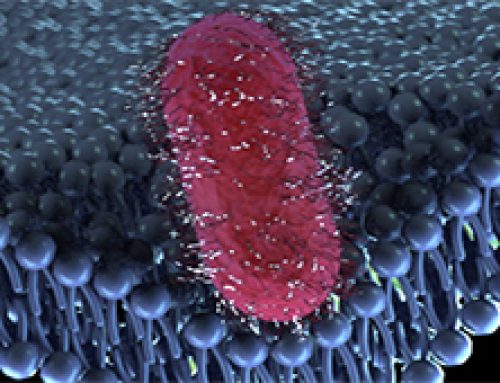
After bacteria enter the host cell, they often form vacuoles that enable them to survive, reproduce, and spread infection. This occurs in both phagocytic and non-phagocytic host cells. The Salmonella Containing Vacuole (SCV) is integrated with the early endocytic pathway, but these vacuoles are able to escape lysosomal fusion and lysis. During SCV maturation, an F-actin meshwork is formed around bacterial vacuoles in a process known as vacuole-associated actin polymerization (VAP) that reinforces the integrity of the vacuolar membrane. Mature SCVs are found in a perinuclear position, proximal to the Golgi apparatus. Salmonellae within SCVs also induce the formation of tubular aggregates along a scaffold of microtubules called Salmonella-induced filaments (SIFs) that extend from SCVs throughout the cell. Therefore, an intricate link exists between the host cytoskeleton and Salmonella pathogenesis at various stages. Other intracellular bacteria like Mycobacteria, Coxiella, Legionella, and Brucella also reside within vacuoles and exploit different components of the endocytic and secretory pathways for pathogenesis.
The Study in Detail
Key Findings
- SsrB regulates biofilm formation independent of SsrA. This was established using ssrA and ssrB null mutants- biofilm levels in ssrB null strain decreased by 60% compared to the ssrA mutant or wild type
- Biofilm formation requires unphosphorylated SsrB. Although phosphorylation of SsrB by SsrA is required for the activation of virulence genes encoded by the SPI-2 pathogencity island, this study shows that phosphorylation of SsrB is not required for biofilm formation. This was demonstrated using an ssrB mutant which was phosphorylation incompetent, strain D56A. D56A mutant could form biofilms similar to wildtype strain, but was avirulent.
- When SsrB is not phosphorylated by SsrA kinase, SsrB binds and bends DNA. The DNA bending is energetically unfavourable for binding by the silencer H-NS which forms rigid nucleoprotein filaments. Thus SsrB binding relieves repression of CsgD, the regulator of biofilm coding genes, and activates its transcription.
Methods and Controls used in the study
- In this study, Scanning Electron Microscopy (SEM) was employed to analyze bofilm morphology
- Virulence of different strains were measured using beta-galactosidase activity of a sifA-lacZ chromosomal fusion
- Atomic Force Microscopy (AFM) imaging was employed to visualize SsrB and H-NS DNA binding.