Project Description
A novel paradigm for the bacterial response to acidic environments in vitro and in a host describes how gram negative bacteria such as Salmonella and E. coli self-acidify for prolonged periods in response to acidic environments resulting from acid or osmotic stress. This is in contrast to previously established views that E. coli rapidly recovers from acidic stress by neutralizing its cytoplasm.
These findings were published in Nature Communications in 2017
Chakraborty S, Winardhi RS, Morgan LK, Yan J and Kenney LJ. (2017) Non-canonical activation of OmpR drives acid and osmotic stress responses in single bacterial cells. Nature Communications, 8:1587. doi:10.1038/s41467-017-02030-0
More information on the Kenney lab and the Yan Lab
Figure: Self-acidification of Salmonella and E.coli in response to acid and osmotic stress: In response to acidic environment resulting from either acid or osmotic stress, EnvZ, the membrane bound sensor kinase is activated by increased helicity of a region surrounding the phosphorylated histidine. Activated EnvZ physically interacts with OmpR to promote OmpR dimerization and DNA binding. In both Salmonella (A) and E.coli (B), OmpR represses the cadC/BA operon in response to acid stress to maintain an acidic cytoplasm. In response to osmotic stress, OmpR represses rpoS, which leads to up-regulation of the oxidoreductase yghA in Salmonella (C), leading to cytoplasmic acidification. In E.coli, the repression of speF which codes for ornithine decarboxylase helps maintain acidic cytoplasm under osmotic stress (D).
This work re-examines the response of Salmonella and E. coli to acid and osmotic stress using sensitive pH measurements by imaging single cells. The results reveal that both Salmonella and E.coli self-acidify their cytoplasm in response to both acid and osmotic stress, and intracellular acidic pH is maintained for prolonged periods. The molecular regulator of self-acidification in both bacteria is OmpR, the key regulator of the EnvZ-OmpR two-component bacterial signaling system. However, the mechanism of OmpR-mediated bacterial self-acidification is different from previously described mechanisms of EnvZ-OmpR signaling through phosphorylation. EnvZ, the membrane bound sensor kinase undergoes activation through increased helicity of the region that surrounds phosphorylated histidine. Activated EnvZ physically interacts with OmpR, promoting OmpR dimerization and DNA binding. OmpR then represses the CadB/A system to maintain acidic internal pH in both Salmonella and E. coli in response to acid stress. In response to osmotic stress, which also causes acidification of the cytoplasm, but to a lesser extent compared to acid stress, the genes OmpR represses are different. In Salmonella, OmpR represses rpoS, which leads to up-regulation of the oxidoreductase yghA. In E.coli, OmpR represses the ornithine decarboxylation (speF) to maintain an acidific cytoplasm.
Understanding the basics
What are two component regulatory systems (TCRS)? Bacteria respond to changing environments by altering gene expression. Two-component regulatory systems (TCRS) are important mediators of signal transduction that enable bacteria to detect physical and/or chemical changes and then relay this signal through the cytoplasm to the bacterial nucleoid, where modulation of gene expression occurs. In its most basic form, a TCRS consists of a membrane-bound sensor kinase and a DNA-binding response regulator. In response to specific stimuli, the sensor kinase is phosphorylated at a conserved histidine residue and then the phosphoryl group is transferred to the conserved aspartate on the response regulator. Phosphorylation of the response regulator triggers a conformational change, driving dimerization and high affinity DNA binding. For example, the SsrA sensor kinase in Salmonella is activated by low pH and phosphorylates the response regulator SsrB. Phosphorylated SsrB de-represses H-NS silencing and activates a set of virulence genes located at the Salmonella Pathogenicity island-2 (SPI-2) region. These genes encode various components of a molecular syringe (Type-III secretion system) which allows the delivery of ~30 specialized effector proteins that manipulate the host cellular machinery to ensure the survival and proliferation of intracellular Salmonella.
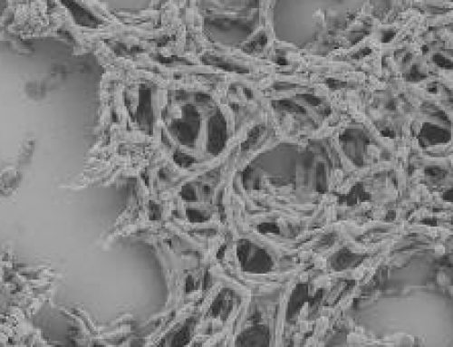
After bacteria enter the host cell, they often form vacuoles that enable them to survive, reproduce, and spread infection. This occurs in both phagocytic and non-phagocytic host cells. The Salmonella Containing Vacuole (SCV) is integrated with the early endocytic pathway, but these vacuoles are able to escape lysosomal fusion and lysis. During SCV maturation, an F-actin meshwork is formed around bacterial vacuoles in a process known as vacuole-associated actin polymerization (VAP) that reinforces the integrity of the vacuolar membrane. Mature SCVs are found in a perinuclear position, proximal to the Golgi apparatus. Salmonellae within SCVs also induce the formation of tubular aggregates along a scaffold of microtubules called Salmonella-induced filaments (SIFs) that extend from SCVs throughout the cell. Therefore, an intricate link exists between the host cytoskeleton and Salmonella pathogenesis at various stages. Other intracellular bacteria like Mycobacteria, Coxiella, Legionella, and Brucella also reside within vacuoles and exploit different components of the endocytic and secretory pathways for pathogenesis.
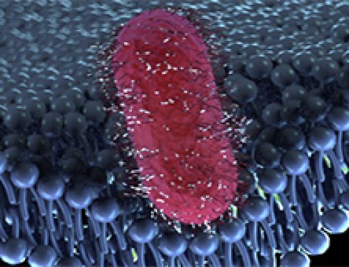
Bacterial cytoplasmic pH can be measured in single cells using the I-switch biosensor labeled at its 5′ and 3′ termini with Alexa-488 and Alexa-647, two different fluorescent molecules. Bacteria are electroporated with the I-Switch The I–switch consists of cytosine-rich unpaired regions that form an anti-tetraplex CH+.C by alternate Watson and Crick base pairing in the presence of protons. This leads to a “closed” conformation of DNA, enabling FRET to occur between the fluorophore pairs. This process is reversible, and at neutral pH, the I-switch dissociates into an open, extended conformation because of electrostatic repulsion between the duplex arms. The FRET intensities provide a measure of the cytoplasmic pH in bacteria.
The Study in Detail
Key Findings
- OmpR regulates acidification of both Salmonella and coli.
- In response to acid stress, OmpR represses lysine decarboxylation regulated by the cadC/BA The repression of cadC/BA prevents the elimination of protons from the bacterial cytoplasm, thus maintaining acidic pH.
- Increasing external osmolality also caused OmpR-dependent acidification in both Salmonella and coli. However, the osmotic stress response is different from acid stress response.
- In Salmonella, OmpR represses the rpoS gene in response to acid stress. Microarray experiments revealed that the self-acidification pathway by repression of rpoS involved yghA, a putative oxidoreductase. YghA is predicted to oxidize NADH to liberate H+, leading to cytoplasmic acidification.
- In coli, OmpR represses the gene encoding ornithine decarboxylase (speF) to prevent recovery from acidification post osmotic stress
- Acidification of Salmonella and coli did not require OmpR phosphorylation. Instead, OmpR was activated by EnvZ through a non-canonical pathway: EnvZ, activated by increased helicity of a region surrounding the phosphorylated histidine, physically interacts with OmpR to promote OmpR dimerization and DNA binding.
- The study establishes that bacterial pH measurements by imaging single cells carrying the pH sensor I-switch or BCECF-AM provides accurate results. It shows that previous methods of pH measurements in bacteria using pHluorin were not accurate as the measurements were averages from a heterogeneous population of bacteria and relied on inefficient clamping agents such as sodium benzoate.
Applications and Future Directions
The finding that gram-negative bacteria such as Salmonella and E. coli self-acidify in response to the acidic host environment as a survival and virulence mechanism opens up novel therapeutic strategies for bacterial infections by preventing bacterial self-acidification.