MBInfo‎ > ‎Modules‎ > ‎

Functional Module: Myosin-X in the Transport of Cargo along Actin Filaments


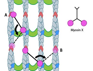
Functional Module: Myosin-X in the Transport of Cargo and Filopodia Initiation

Filopodia are motile structures that contribute to the cell’s ability to detect mechanical or chemical signals and measure and respond to its physical environment. This mechanosensing is mediated primarily by adhesions and receptor molecules located at the tips, or along the shafts, of filopodia.

The transport of components along filopodial shafts is crucial to the continued growth of actin filaments and the formation of adhesions at the tips of filopodia. Myosin-X facilitates the transport of components from the cell body to filopodial tips, resulting in their integration into the cell membrane as receptors/adhesions or their use in the growth of actin filaments and filopodia [1, 2, 3, 4, 5].

Movement

Movement of myosin-X is driven by ATP hydrolysis, in a unique mechanism that resembles walking or stepping. This movement is known to occur preferentially on actin bundles rather than single actin filaments [6, 7]. Although it is essentially a forward movement, evidence indicates that the protein may also take side-steps. This may be carried out as a means of overcoming obstacles or defects in the track [7].

The walking mechanism of myosin-X is known to be distinct from other walking myosins such as myosin-V. This is particularly relevant to the step size and the preference for specific actin bundling proteins [7]. Whilst myosin-V takes steps of 36nm and follows single filaments rather than side-stepping along parallel filaments of the bundle, myosin-X takes shorter steps which have been measured at 17.5nm [6, 7]. This shorter stepping distance may explain why myosin-X uses binding sites on parallel filaments, as a short step on a single filament would result in the protein rotating around the natural twist of the filament [7].


Figure: Myosin-X step size corresponds to a single twist of the actin filament helix. Binding sites are represented by red and dark blue actin monomers. In this figure we see how Myosin-X is able to step forwards and backwards (A) as well as horizontally, between filaments (B).

Cargo Binding

Myosin-X is able to carry a variety of cargo to the tips of filopodia [1, 2, 3, 4]. Although the exact mechanism by which myosin-X selects cargo remains unclear it is well established that cargo recognition and binding results from the presence of the myosin tail homology 4 (MyTH4) and ezrin/radixin/moesin (FERM) domains – together these are known as the MyTH4-FERM cassette [8].

A number of components essential to the growth and function of filopodia are carried by myosin-X, including β-integrin [3]. β-integrin is essential for focal adhesion formation and binds to myosin-X via the FERM domain. Knock down of myosin-X by siRNA was shown to disrupt the formation of adhesions to collagen I, particularly at early time points, with no disruption after 60 minutes. This highlights the importance of myosin-X in the initial stages of integrin-mediated adhesion formation [3].

Other cargo transported by myosin-X includes neogenin and ‘deleted in colorectal cancer’ (DCC), which function as netrin receptors. In addition, DCC anchors translational machinery such a ribosomes to the membranes of neuronal growth cones and dendrites [8, 4].

A Role in Filopodia Initiation and Elongation

As well as mediating cargo transport along actin filament bundles, recent studies have implicated myosin-X as integral to the initiation of filopodia and the elongation of long filopodia.

Studying the localization and motility of single myosin-X molecules using TIRF microscopy, Watanabe et al, hypothesized that binding of cargo, and the preceding dimerization of myosin-X monomers at the cell periphery is important for filopodia initiation as it promotes actin filament bundling [9]. This was proposed to occur in a similar manner to regular crosslinking. Previous reports had indicated that even without the cargo-binding FERM domain, lateral movement of myosin-X along the leading edge of lamellipodia promoted actin reorganization and through its motor activity, filopodia initiation. In this case the length of the head and neck domain of myosin-X was proposed to be important for initiation. In the study by Watanbe et al a rapid increase in the rate of recruitment and assembly of myosin-X at filopodia initiation sites moments before protrusion commenced was observed and again this was shown to be independent of the presence of the FERM domain.

After removing the FERM domain from myosin-X (using a FERM domain-truncated construct called M10-ΔFERM) the protein was still able to walk along actin filament bundles and continued to localize at both the leading edge and filopodia tip. Significant changes to the length and stability of the growing filopodia were however noted. Not only were filopodia significantly shorter and more unstable, as previously reported [5, 3], but the phased-extension mechanism of elongation observed when complete myosin-X was present, did not occur.

Although it was noted that without the FERM domain the transport of essential cargo to the tips of the filopodia would be insufficient to permit continued elongation, it was also proposed that through FERM-
β-integrin interactions at the tips of filopodia (when filopodia are attached to substrates via focal adhesion sites), myosin-X may also possess adhesive qualities. In this hypothesis, as filopodia enter the retraction phase and actin filaments move back towards the cell body by retrograde flow, myosin-X remains at the tips of filopodia together with the adhesion complex. Upon shrinkage of the tips, the protein will rebind to actin filament bundles and allow a new phase of extension to begin from adhesion sites. This mechanism of phased-extension is supported by observations that without the FERM domain myosin-X diffuses back into the cell body during the retraction phase whilst intact myosin-X remains at the tips [9].

Č
Ĉ
ď
Steven Wolf,
Dec 12, 2011, 7:39 PM
Ĉ
ď
Steven Wolf,
Dec 12, 2011, 7:39 PM
Comments