
Additional Links
Essential for: Initiation in Filopodia Formation Initiation in Lamellipoda and Lamella Focal adhesion initiation Podosome extension Related in Function to: Arp2/3 Mediated Nucleation | Functional Module: Formin and Profilin in Actin NucleationActin filament assembly and disassembly are primary molecular processes that facilitate whole cell motility and the movement of subcellular structures. The initial stage in the formation of actin filaments is nucleation. This is defined as the formation of a stable actin polymer from its monomeric units [1].Although nonmuscle cells have a high concentration of G-actin-ATP (~100 μM) [2], pure G-actin monomers fail to nucleate new actin filaments efficiently due to the instability of actin oligomers. Additional factors are therefore necessary for the production of actin filaments. Several models of actin nucleation have been described [1], including formin-mediated nucleation, which involves a number of proteins, particularly members of the formin family, profilin and G-actin. Members of the formin family of proteins (that includes mDia) are key regulators of unbranched actin filament initiation and nucleation (reviewed in [3]). They are essential for generating, extending and/or protecting lamellipodial actin filaments from capping [4] and are also also key regulators of actin bundle initiation and formation in filopodia [5, 6] and stress fibers [7, 8, 9]. The formin, mDia2, is suggested to be involved in both initiation of invadopodia formation through actin nucleation and subsequent growth of invadopodia through the elongation of actin filaments [10]. Formins are essential in the nucleation of new filaments, as they stabilize actin dimers (reviewed in [11]). Under normal circumstances they are auto-inhibited through structural interactions between the two ends of the protein [12]. However, conformational rearrangements resulting in their activation can be induced through interactions with GTP-bound (active) Rho GTPases [13]. This process remains poorly understood (reviewed in [3]). Latest Findings In a recent study, the tumor suppressor adenomatous polyposis coli (APC) was shown to bind the formin mDia1 and overcome capping protein- and profilin-mediated suppression of spontaneous actin nucleation, resulting in the initiation of actin filament nucleation and elongation [14]. In the mechanism described, APC is primarily responsible for actin monomer recruitment, whilst mDia1 catalyzes filament elongation. Actin recruitment by APC did not involve capturing F-actin intermediates that had spontaneously formed nor did APC contribute to filament elongation. In this model, once actin polymerization commences the APC-mDia1 complex separates – mDia1 is propelled away from APC along with the growing barbed end of the filament and APC remains attached to the filament at the site of nucleation [14]. Although a consensus has yet to be reached for the mechanism of formin-mediated nucleation, it is now well-established that activated formins function as dimers and form a donut-shaped complex around terminal actin subunits, orientating themselves toward the (+) end of the actin filament or nucleus [15]. This binding is facilitated by FH2 (formin homology 2) domains within the formin monomers. Next, each formin monomer binds and captures profilin units, which are themselves already bound to G-actin monomers. This interaction is mediated by multiple stretches of polyproline residues within the FH1 domain of formins [16]. This domain is known to range from 15-229 residues, consist of between 35% and 100% proline residues, and contain up to 16 profilin binding sites [17]. Profilin maintains a steady pool of actin monomers by promoting ADP to ATP nucleotide exchange on G-actin [15]. These monomers of ATP-G-actin are then added the growing actin filament. The coupling of formin with the growing end prevents capping and allows continued growth of the filaments [18]. 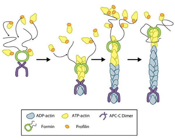 Figure: Formin-mediated nucleation of actin filaments. The FH2 domains of the formin dimer (shown in green) bind to actin monomers to initiate filament assembly. Recent studies indicate this is assisted, or even mediated, by additional factors such as APC. The FH1 domains of the formin dimer (shown as black lines) have short polyproline sequences that interact with profilin. Profilin binds to both formin and actin monomers to increase the addition of actin monomers to the barbed end of the filament. |
|
|
|
Current MCMF Content |
Functional Module: Formin and Profilin in Actin NucleationČ |
MBInfo © 2013 National University of Singapore.
MBInfo is sponsored by:
Sign in|Report Abuse|Print Page|Remove Access|Powered By Google Sites