Focal Adhesion Initiation

Cell-Matrix Adhesions

5.4 Initiation of Focal Adhesion Assembly

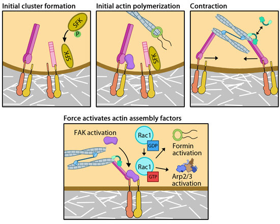
Focal adhesion formation is initiated upon the binding of adhesion receptors to extracellular matrix (ECM) ligands (e.g. fibronectin, vitronectin, collagen) along the cell periphery usually at the protruding edge of a cell. Both intracellular and extracellular factors can influence the level of matrix binding, in terms of affinity (the strength of interactions, reviewed in [1, 2]) and avidity (the number of interactions, such as lateral interactions between independently activated proteins within a focal adhesion). Nascent focal adhesions first appear exclusively in the lamellipodium as submicron-sized puncta that are typically immobile but can at times travel short distances along the direction of the actin retrograde flow [3, 4]. 


As the primary ECM receptor in the FAs, integrins are heterodimeric transmembrane protein, with large multidomain extracellular portions and small cytoplasmic tails. Although there are many different types of integrins with specificity to different ECM, a major portion of cellular and biophysical studies have focused on fibronectin-binding α5β1 and αvβ3 integrins. β1 integrins have been shown to exhibit catch bond behavior [5, 6] and function as the force-bearing component. In this capacity β1 integrins maintain adhesion strength despite fluctuating matrix forces, which can often change quite rapidly. However, it is unclear whether β1 integrins bind matrix molecules at the leading edge and translocate inwards [7, 8] or get recruited at later stages [9, 10]. The less stable β3 integrins are responsible for initiating mechanotransduction and reinforcing FA attachments to the ECM, in complex with talin [11].
Existence of a phosphorylation-dependent crosstalk between the two integrin types during migration has also been reported [12].

Upon binding of ligands by integrins and clustering, a number of signal transduction cascades are activated as described
below. One key event is Rac1 activation and consequent phosphoinositide production [13, 14], which leads to the recruitment of talin homodimer [15, 16]. For further details, see Functional Module: Integrin β1/syndecan-4 synergy in adhesion dynamics. Recent studies provide evidence that anchoring of talin at FAs also requires F-actin and vinculin [17]. This is followed by integrin recruitment and activation upon fibronectin binding [18]. The talin-mediated linkage to the actin cytoskeleton serves to stabilize the integrin-ECM bonds [7]. As talin is pulled by the moving actin, it either stretches leading to rearward translocation of β1 integrins and unfolding of fibronectin [19] or results in frequent slip bonds of 2 pN on immobilized β3 integrins [20, 21]. Thus a dynamic nanoscale organization of integrins exists inside FAs, determined by their extracellular domains [18]. Further, this study also reports a distinct dynamics of integrins within FAs, where they constantly alternate between ligand-bound activated and unbound inactivated states, which is thought to confer the adaptability to FAs in order to withstand rapid changes in force [22].

Talin
is one of the best characterized FA proteins that play important roles during FA initiation. In addition to its binding site to integrin cytoplasmic tails that can activate integrin (reviewed in [1]), talin can also bind directly to actin and associate with numerous cytoskeletal and signaling proteins (reviewed in [23]), effectively forming one of the core cell-ECM mechanotransduction units.

With the integrin considered ‘engaged’, the adhesion complexes are capable of exerting low level forces to fibronectin, leading to a cascading series of events: 


1) the rearrangement of ECM ligand domains to adjust the length of talin

2) the strengthening of the talin-actin slip bond [20]
3) the slowing down of the actin retrograde flow which also helps to prevent the disintegration of the nascent adhesion complexes [3]
.


In addition to integrins, syndecan-4 also plays an essential role [24, 25], binding different domains of matrix proteins and eliciting cooperative signals (reviewed in [26]). Several membrane proteins are also believed to be interacting with integrins, likely as co-receptors, although their functions in cell migration and mechanotransduction remain not well understood (reviewed in [27]).

It should be noted that nascent adhesions do not uniformly undergo these sequential growth. Rather, 
shear forces generated by actin retrograde flow result in the disassembly of certain fraction of nascent adhesions, and only a subset of nascent adhesions survive to proceed into later stages.

Integrin Clustering, Cytoskeletal Linkage and Signal Transduction

There is still much debate over the mechanisms of signal transduction following integrin-ligand binding, however recent studies reveal that irrespective of the global density of integrins, local clustering of ligand-bound integrins is paramount to efficient signal transduction [28].
Latest Findings  The minimum cluster area required for stable FA assembly and force transmission has a dynamic nanolimit, regulated by the interplay between adhesive force, cytoskeletal tension and the structural linkage that transmits them [29]. These initial clusters (specifically αVβ3 integrins) serves as a platform for the tethering and polymerization of actin filaments [30, 31]. For details on actin filament assembly, see Functional Module: Formin and Profilin and Functional Module: Arp2/3 complex.

From available data, the following course of events is hypothesized to be the working model for FA initiation (reviewed in [32], see figure below).

Actin-talin-integrin complexes stabilize both focal adhesions and stress fibers through the recruitment of additional components such as focal adhesion kinase (FAK) [33, 34], paxillin [35, 36] and Src-family kinases (SFKs) to integrin tails [37, 38]. The phosphatase, RPTP-α, is known to activate SFKs and in doing so reinforce αVβ3-cytoskeletal connections [39, 40]. While talin can further enhance PIP2 production and FAK activation [41, 42], FAK has been shown to reinforce clusters by promoting talin recruitment [43]. Src-dependent actin polymerization initially pushes the clusters outwards.

Subsequently, myosinII contraction brings adjacent actin-linked clusters closer to one another and inwards [31]. Myosin contractility on attached actin filaments has also been shown to stretch talin [44], revealing binding sites for other proteins, such as vinculin [45, 46, 47]. Thus it is believed that cycles of talin deformation, vinculin binding and release by the slipping actin filaments [25] integrate the traction exerted on the substrate to biochemical signals [44].

Figure: Events leading to focal adhesion initiation. Adapted from [32]. Initial integrin clusters (top left), after activation by talin binding, provide avenue for initial actin polymerization (top middle) by recruiting focal adhesion components- FAK, SFKs and paxillin. New actin filaments tether to talin, the clusters get pushed away and then pulled closer by myosin contractions (top right). This causes cycles of transient talin stretching and vinculin binding until the talin- actin bond stabilizes. Upon stable vinculin binding (bottom), further integrin clustering and signaling promote Rac1 activation as described elsewhere. Rac1, in turn, further activates actin polymerization modules, Arp2/3 and formins.


Once the talin-actin bond stabilizes, the talin-bound vinculin augments actin coupling through its tail [48] and promotes allosteric integrin clustering via signal transduction molecules such as Rap1 GTPase (reviewed in [8, 49]). Actin polymerization is also believed to precede adhesion formation, for example through the direct interaction of vinculin with components of the actin polymerization module such as VASP [50, 51] and Arp2/3 in a PI3K and Rac dependent manner [52, 53]. Although the mechanism of filament nucleation at adhesions is elusive, several Rac1 targets are implicated (reviewed in [54]). 


Additionally, the link between the actin cytoskeleton and integrins can also be further stabilized by the recruitment of the IPP cytosolic ternary complex [55], comprising integrin-linked kinase (ILK), parvin and PINCH (particularly interesting Cys-His-rich protein). Integrin-related kinase (ILK) [56] is able to bind to the separated tails of activated β1 and β3 integrins [57, 58]. The IPP complex is recruited to focal adhesions to promote cytoskeleton linkage and integrin signaling [58, 59] through several binding partners including paxillin and kindlin (reviewed in [60]).
Č
Ĉ
ď
Steven Wolf,
Jan 9, 2012, 8:13 PM
Ĉ
ď
Steven Wolf,
Jan 9, 2012, 8:22 PM