
Read Further…
Phosphoinositides are required in the activation of: Talin; Vinculin; alpha-actinin; Focal adhesion kinase; Nucleation promoting factors
They are also required for the inhibition of: ADF/ cofilin (AC) family; Capping protein
Essential for: Actin assembly; Focal adhesions initiation; Syndecan dimerization and signaling
They are also required for the inhibition of: ADF/ cofilin (AC) family; Capping protein
Essential for: Actin assembly; Focal adhesions initiation; Syndecan dimerization and signaling
Phosphoinositides
are a major component and building block of most biological cell
membranes. Phosphoinositides have amphipathic behavior, meaning they
contain two segments that differ in their biochemical properties: the
first segment is two hydrocarbon “tails” which are nonpolar or
hydrophobic which causes them to move away from water; the second
segment is the “head group” or inositol ring, which is polar or
hydrophilic, thus allowing it to interact with water. Because a glycerol
backbone connects the two segments, the phosphoinositides are grouped
into a broader category of membrane lipids known as phosphoglycerides.
The core component of a phosphoinositide is called phosphatidylinositol
(see “Figure: General structure of phosphoinositides” below).
Three main types of phosphoinositides have important roles in intracellular signaling, lipid signaling, and membrane trafficking; these phosphoinositides differ solely in the number of phosphate groups that are attached by phosphoinositol kinases to the inositol ring (see “Figure: Phosphoinositides involved in cell signaling” below):
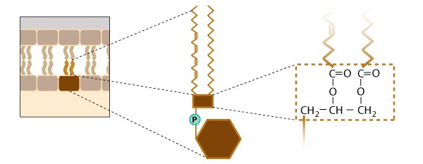
Figure: General structure of phosphoinositides [1]. The composition and length of the hydrocarbon tail is somewhat diverse and is reported to range from 16 carbons to 22 carbons. For simplicity, the glycerol backbone in this illustration is represented by a brown box, and the oxygen atoms of the inositol head group have been omitted. Chemical modification to the inositol ring occurs in the conversion of phosphatidylinositol (shown here) to other phosphoinositides (see figure below).

Figure: Phosphoinositides involved in cell signaling. This figure gives an example of how different phosphoinositides are produced from phosphatidylinositol by the action of two kinases (e.g., PI kinase and PIP kinase) and it shows how signaling molecules (e.g. PIP2, DAG, IP3) are generated. It should be noted that a number variations exist for which hydroxyl groups are phosphorylated, and not surprisingly, a number of kinases are involved in these phosphorylation events. DAG= diacylglycerol; IP3=inositol triphosphate.
Three main types of phosphoinositides have important roles in intracellular signaling, lipid signaling, and membrane trafficking; these phosphoinositides differ solely in the number of phosphate groups that are attached by phosphoinositol kinases to the inositol ring (see “Figure: Phosphoinositides involved in cell signaling” below):
- Phosphatidylinositol-4-phosphate (PIP)– increased levels of PIP in the plasma membrane greatly reduces the F-actin binding and depolymerizing activity of ADF (actin depolymerizing factor).
- Phosphatidylinositol-4,5-bis-phosphate (PIP2) – increased levels of PIP2 in the plasma membrane inhibits actin filament capping by capping protein and greatly reduces the F-actin binding and depolymerizing activity of ADF.
- Phosphatidylinositol-3,4,5-trisphosphate (PIP3) – Phosphatidylinositol-3-kinase (PI3K) and PTEN (Phosphatase and tensin homolog) signal transduction pathways regulate the level of PIP3 in response to extracellular guidance cues during filopodia motility. The accumulation of PIP3 in filopodia is suggested to cause actin polymerization and increased cellular movement.
Figure: General structure of phosphoinositides [1]. The composition and length of the hydrocarbon tail is somewhat diverse and is reported to range from 16 carbons to 22 carbons. For simplicity, the glycerol backbone in this illustration is represented by a brown box, and the oxygen atoms of the inositol head group have been omitted. Chemical modification to the inositol ring occurs in the conversion of phosphatidylinositol (shown here) to other phosphoinositides (see figure below).
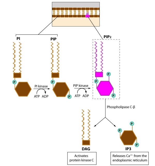
Figure: Phosphoinositides involved in cell signaling. This figure gives an example of how different phosphoinositides are produced from phosphatidylinositol by the action of two kinases (e.g., PI kinase and PIP kinase) and it shows how signaling molecules (e.g. PIP2, DAG, IP3) are generated. It should be noted that a number variations exist for which hydroxyl groups are phosphorylated, and not surprisingly, a number of kinases are involved in these phosphorylation events. DAG= diacylglycerol; IP3=inositol triphosphate.