Sarcomeres facilitate contractility of skeletal muscle[Edit]
A Role in Contraction Numerous motor proteins exist and each possess unique characteristics that allow them to facilitate different processes and functions. Even within the myosin superfamily variation exists in structure and function of each member. Not only can motor proteins translocate along microfilaments, but they can induce movement in the filament itself. It is this property that gives rise to the contractile properties of skeletal muscle.
 Figure 1. Actin-myosin contraction in muscle cellsIn skeletal muscle cells, myosin II forms only thick filaments that are arranged within a scaffold of actin thin filaments (along with numerous other proteins). These form the higher order fibrous structures known as sarcomeres. Each sarcomere contains numerous repeating units of interlinked thick and thin filaments, and the opposite orientation of the myosin heads causes adjacent actin filaments to slide past each other during muscle contraction. Each sarcomere is ~2 µm long in resting muscle, but this length is shortened by as much as 70% after muscle contraction. Muscle contraction is regulated by calcium levels [1] and by the troponin regulatory system. Although actin subunits continue to turn-over at both ends of the thin filament, this exchange is relatively slow, making the actin filaments in sarcomeres relatively more stable when compared to the actin filaments found in other cell types.
Video: Sliding filament theory of muscle contraction. [Video was uploaded to YouTube by avice01 and created by Sara Egner using electron micrography from P M Motta, P M Andrews, K R Porter and J Vial.]
Contractile Bundles in Skeletal Muscle
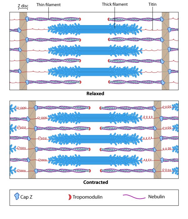 Figure 1. Actin-myosin contraction in muscle cells