Topics

    What is axon guidance and the growth cone?[Edit]

    Axon guidance is an important step in neural development. It allows growing axons to reach specific destinations and ultimately form the complex neuronal networks throughout the body. Although many aspects of this mechanism remains unclear, it is well established that a dynamic and highly motile actin-based structure found at the growing end of a developing axon, known as a growth cone, facilitates this process. 

    The Growth Cone
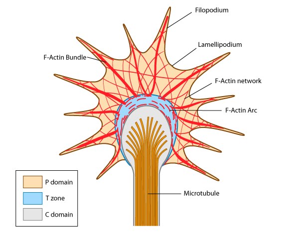
    Figure 1. Growth cone structure
    Growth cones facilitate axon growth and guidance by bundling and extending actin filaments into structures known as filopodia and microspikes. Binding of filopodia and adhesion receptors to particular extracellular matrix (ECM) components or ligands is translated into actin filament assembly, cytoskeleton remodeling and force-driven motility. These events culminate in the growth of the neuron towards its target. 

    Growth cones contain a number of cytoskeletal components that are organized into three regions; the peripheral (P) domain, the transitional (T) domain and the central (C) domain [1]:

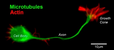
    Figure 2. CD1 mouse spinal commissural neuron with growth cone
    The P domain is primarily composed of unipolar actin filament bundles embedded in a less polar actin network. It contains dynamic lamellipodia and filopodia. Microtubules are also transiently found within this domain. 

    The C domain is located in the center of the growth cone nearest the axon. It is primarily composed of microtubules and contains numerous organelles and vesicles. 

    The T domain is a thin interface between the C and P domains.

    A number of cytoskeletal-associated proteins are present in growth cones that anchor actin filaments and microtubules to each other (e.g. myosin II [2]), to the membrane (e.g. talin [3]) and to other cytoskeletal components. Molecular motors present in growth cones produce the forces needed for growth cone migration (e.g. myosin II [4]) and vesicle transport in and out of the growth cone (e.g. KIF4 (kinesin superfamily protein member 4) [5]).

    References

    1. Suter DM., Forscher P. An emerging link between cytoskeletal dynamics and cell adhesion molecules in growth cone guidance. Curr. Opin. Neurobiol. 1998; 8(1). [PMID: 9568398]
    2. Burnette DT., Ji L., Schaefer AW., Medeiros NA., Danuser G., Forscher P. Myosin II activity facilitates microtubule bundling in the neuronal growth cone neck. Dev. Cell 2008; 15(1). [PMID: 18606149]
    3. Sydor AM., Su AL., Wang FS., Xu A., Jay DG. Talin and vinculin play distinct roles in filopodial motility in the neuronal growth cone. J. Cell Biol. 1996; 134(5). [PMID: 8794861]
    4. Peretti D., Peris L., Rosso S., Quiroga S., Cáceres A. Evidence for the involvement of KIF4 in the anterograde transport of L1-containing vesicles. J. Cell Biol. 2000; 149(1). [PMID: 10747093]
    5. Medeiros NA., Burnette DT., Forscher P. Myosin II functions in actin-bundle turnover in neuronal growth cones. Nat. Cell Biol. 2006; 8(3). [PMID: 16501565]
    Updated on: Mon, 20 Oct 2014 09:38:22 GMT