Myosin motor protein structure[Edit]
Myosin is a motor protein that uses the energy from ATP hydrolysis to move along actin filaments. Many myosin isoforms have been found in eukaryotes (See Figure below), which differ in the type of heavy and light chains they are composed of. All myosins are composed of a diverse ‘tail’ domain at their carboxy terminus and an evolutionarily conserved globular ‘head’ domain at their amino terminus.
 Figure 1. The myosin superfamily of motor proteins: All myosins share a motor domain on their heavy chains at the amino-terminus (the ‘head’ domain), but they differ considerably at their carboxy-terminus (the ‘tail’ domain). A few myosin types also have an amino-terminal extension. The number of light chains varies considerably between myosin types and certain myosins exist as dimers. Myosins that form dimers have two motor domains, and the number of light chains can influence the “lever arm” length between the myosin heads – this regulates the length of the myosin ‘powerstroke’ and the distance the myosin can travel along the actin filament in a single round of ATP hydrolysis (see also ‘myosin powerstroke’).
The diverse ‘tails’ of different myosin isoforms bind specific substrates or cargo, whilst their conserved ‘heads’ contain sites for ATP binding [1], F-actin binding and force generation (i.e. motor domains) (reviewed in [2, 3]).
Figure 1. The myosin superfamily of motor proteins: All myosins share a motor domain on their heavy chains at the amino-terminus (the ‘head’ domain), but they differ considerably at their carboxy-terminus (the ‘tail’ domain). A few myosin types also have an amino-terminal extension. The number of light chains varies considerably between myosin types and certain myosins exist as dimers. Myosins that form dimers have two motor domains, and the number of light chains can influence the “lever arm” length between the myosin heads – this regulates the length of the myosin ‘powerstroke’ and the distance the myosin can travel along the actin filament in a single round of ATP hydrolysis (see also ‘myosin powerstroke’).
The diverse ‘tails’ of different myosin isoforms bind specific substrates or cargo, whilst their conserved ‘heads’ contain sites for ATP binding [1], F-actin binding and force generation (i.e. motor domains) (reviewed in [2, 3]).
All myosins bind to actin filaments via a globular ‘head’ domain located at the end of the heavy chains. Actin binding to this region increases the ATPase activity of myosins (reviewed in [2][4]. Some myosins have a single heavy chain and contact actin filaments at only one site, while other myosin isoforms have two heavy chains and contact actin filaments at two sites. Myosin II is the only family member that can form polymeric assemblies[3]) (See “thick filaments” below).
The number of light chains influences the length of the “lever arm” or “neck region” and therefore the “step size” of different myosin types [5]. Myosin V contains more light chains relative to myosin II and so myosin V moves in larger steps along actin filaments after an equivalent round of ATP hydrolysis (reviewed in [6]).
Myosin motors move along actin filaments in defined directions. With the exception of myosin VI, which moves towards the pointed end, all myosins move towards the barbed end. Most actin filaments have the barbed end directed towards the plasma membrane and the pointed end towards the interior. This arrangement allows certain myosins (e.g. myosin V) to function primarily for cargo export, while myosin VI acts as the major motor protein for import. Myosin II is commonly associated with retraction fibers and retrograde actin flow at the pointed end of actin filaments. All non-muscle cells use contractile bundles containing myosin II to generate forces that promote the assembly of actin filaments.
Although most myosins function as motor proteins in the cytoplasm, some species of myosin are localized to, and function in, the nucleus. Nuclear Myosin I (NMI) myosin II, myosin V, myosin VI, myosin XVIB and myosin XVIIIB have all been found in the nucleus [7, 8, 9], with NMI being the most extensively studied.
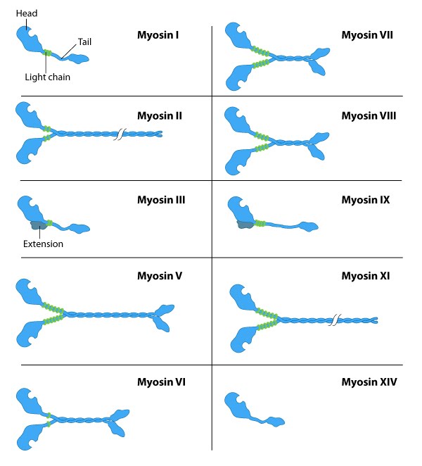 Figure 1. The myosin superfamily of motor proteins: All myosins share a motor domain on their heavy chains at the amino-terminus (the ‘head’ domain), but they differ considerably at their carboxy-terminus (the ‘tail’ domain). A few myosin types also have an amino-terminal extension. The number of light chains varies considerably between myosin types and certain myosins exist as dimers. Myosins that form dimers have two motor domains, and the number of light chains can influence the “lever arm” length between the myosin heads – this regulates the length of the myosin ‘powerstroke’ and the distance the myosin can travel along the actin filament in a single round of ATP hydrolysis (see also ‘myosin powerstroke’).
All myosins bind to actin filaments via a globular ‘head’ domain located at the end of the heavy chains. Actin binding to this region increases the ATPase activity of myosins (reviewed in [2][4]. Some myosins have a single heavy chain and contact actin filaments at only one site, while other myosin isoforms have two heavy chains and contact actin filaments at two sites. Myosin II is the only family member that can form polymeric assemblies[3]) (See “thick filaments” below).
The number of light chains influences the length of the “lever arm” or “neck region” and therefore the “step size” of different myosin types [5]. Myosin V contains more light chains relative to myosin II and so myosin V moves in larger steps along actin filaments after an equivalent round of ATP hydrolysis (reviewed in [6]).
Myosin motors move along actin filaments in defined directions. With the exception of myosin VI, which moves towards the pointed end, all myosins move towards the barbed end. Most actin filaments have the barbed end directed towards the plasma membrane and the pointed end towards the interior. This arrangement allows certain myosins (e.g. myosin V) to function primarily for cargo export, while myosin VI acts as the major motor protein for import. Myosin II is commonly associated with retraction fibers and retrograde actin flow at the pointed end of actin filaments. All non-muscle cells use contractile bundles containing myosin II to generate forces that promote the assembly of actin filaments.
Although most myosins function as motor proteins in the cytoplasm, some species of myosin are localized to, and function in, the nucleus. Nuclear Myosin I (NMI) myosin II, myosin V, myosin VI, myosin XVIB and myosin XVIIIB have all been found in the nucleus [7, 8, 9], with NMI being the most extensively studied.