SIF_AdhesionFormation

Lamellipodia and Lamella

Step 3: Formation of Adhesions

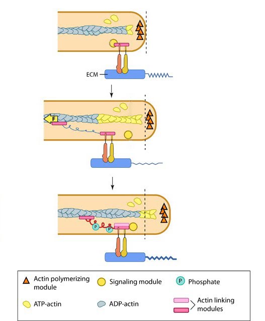
As the lamellipodia continues to spread and expand forward, adhesion sites form along the leading edge (see video below). These sites not only provide traction for the cells forward movement, but permit the cell to sense and measure the rigidity of its substrate. Formation of these adhesions is a complex process, involving a number of steps and functional modules.

To read more about adhesion formation please follow the link to Focal Adhesion Initiation.

Video: Focal adhesions are essential for cell spreading. Upon neuregulin treatment, cell-surface receptors Erb B3/B4 induce lamellipodia formation, most probably through activation of the Rac-WAVE-Arp2/3 pathway. During lamellipodial protrusion, numerous focal adhesions form along the cell periphery and can be visualized as fluorescent spots (GFP-VASP). [Source: Leticia Carramusa, Weizmann Institute of Science, Israel. Permission: Alexander Bershadsky, Mechanobiology Institute, Singapore.]

Particularly important in lamellipodia function is the spatial arrangement of adhesions at the leading edge. Lamellipodia extension and retraction is not uniform across the whole cell and similarly, adhesion sites will not be uniformly distributed. Instead a number of factors may influence arrangement including chemistry of the extracellular matrix, substrate stiffness and cells growth conditions, as described under ‘matrix properties’.

Interestingly, distribution of adhesion sites was found to be coupled to actin polymerization, with conformationally activated, but unliganded β1 integrins, shown to be interacting with elongating filaments [1]. Importantly, this interaction permits a sideways distribution along the leading edge, ensuring clusters of β1 integrin are positioned at the very front of cell protrustions.
β1 integrin was also found, in the same study, to localize at the tips of growth cone filopodia [1].

Focal Adhesions as Molecular Clutches:

Once formed, focal adhesions essentially act as “molecular clutches”, promoting protrusion at the leading edge whilst suppressing membrane contraction (reviewed in [2, 3, 4]).
Adhesions aid forward movement by regulating the forces produced by actin dynamics in different cellular compartments through several methods:

1) They aid membrane protrusion by resisting actin retrograde flow [5] and hence, indirectly promote the force produced by lamellipodial actin polymerization.
2) They convert myosin pulling forces at the lamellar interface into traction forces against the ECM that pulls the cell body forward [6, 7].

The efficiency of this molecular clutch in converting force into protrusion is variable. As some of the adhesion components move along with the retrograde flow [8], the clutch slips [9]. The rate of forward protrusion increases when actin and actin-adhesion linking components become more organized [10] and the retrograde flow of actin is slowed down [5, 11] when the clutch is “partially engaged”. As the adhesions grow and mature under stress, the clutch transforms from being “partially/locally engaged” to “engaged” [2] and hence can influence global cell behavior [12, 13].

To find out more on the forces generated by the within the lamellipodia, read Step 4: Force Generation and Translocation.


Figure: Focal Adhesions act as Molecular Clutch during Forward movement. In the lamellipodium, actin flows backward due to actin polymerization in the front (yellow monomers), depolymerization at the rear (grey monomers) and actomyosin pulling (yellow arrow in middle panel). Top: On binding ECM ligand, proteins are recruited (pink block) to the integrin tails. Middle: These then cluster to provide a dynamic linkage between integrins and the actin filaments, that acts as a clutch. These linkages are weak and could be broken by the retrograde actin flow, hence the clutch is considered partially engaged. Bottom: Under tension, the clutch becomes engaged by linkage reinforcement through phosphorylation events. Force generated by actin dynamics gets converted into traction forces on the matrix molecules (thickening of ligand tail). This also slows down the actin flow and provides hold for the actin polymerization, which pushes the membrane forward.

Č
ċ
ď
LPadhesionformation_proteins.csv
(1k)
Sruthi Jagannathan,
Mar 21, 2012 11:42 PM