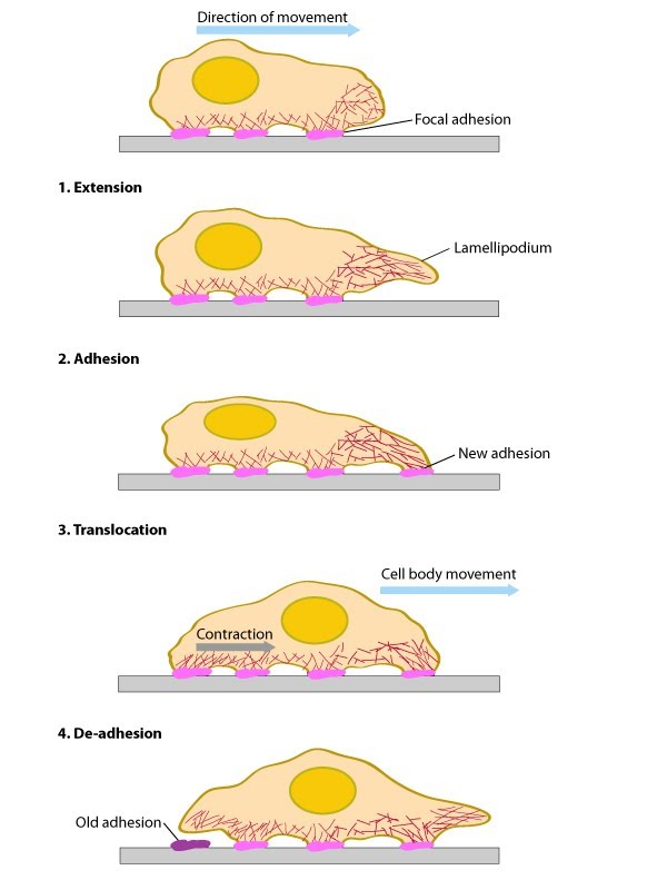
Lamellipodia-Steps in formation: Overview


< PREVIOUS STEP | NEXT STEP >

Lamellipodia and Lamella

Steps in Formation: Overview

In general, cell motility requires two types of forces: 1) a protruding force to extend the leading edge forward; and 2) traction forces to move the cell body [1] (reviewed in [2,3]). These driving forces are functionally-integrated and they are modulated by actin filament dynamics [4,5-9] (reviewed in [10-12]). Cell spreading and motility also require plasticity at the plasma membrane and cell adhesions (reviewed in [13] in addition to transcriptional control of gene expression [14]).

The lamellipodia and lamella are two distinct regions of the cell that facilitate cell motility and function in mechanosensing mechanisms. These regions undergo defined steps in their formation and function as is described in this section of the manual.

In summary, the steps of formation and function are:

  1. Actin filament polymerization produces a protrusive force on the cell membrane that promotes a spreading out and enlargement of the lamellipodia. In polarized, migrating cells this is known as the leading edge.
  2. As new actin filaments form at the leading edge the lamellipodia will continue to extend. Mature filaments are either drawn into a more stable filament network within the lamella, interacting with focal adhesions that reside at the interface between the lamellipodia and lamella.
  3. Focal adhesion formation continues to occur at the leading edge and deeper into the lamella. Adhesion sites may facilitate rigidity sensing and other mechanosensing mechanisms. Through interactions with the substrate, these sites will also provide a means of traction for the cell to pull itself along.
  4. Contractions of the actomyosin network are required for both mechanosensing and cell motility mechanisms. In the former scenario, contractions are required for the cell to pull against its substrate, thereby measuring its rigidity. In migrating cells, contractions are required to translate generated forces into motility, pulling the cell against the focal adhesions and facilitating the retraction of the trailing edge.
  5. The final step in the function of the lamellipoida/lamella is the retraction of the trailing edge. This ensures the cell is not permanently anchored to its substrate and therefore allows a net gain in forward movement
Figure: Conserved steps in cell spreading and movement. 1. Polymerization of actin filaments at the leading edge is translated into protrusive force. 2. Membrane protrusion facilitates the binding of transmembrane cell surface receptors to the substratum components. New adhesions are rapidly linked to the network of actin filaments. 3. The combined activity of retrograde actin movement and contractile forces produced by stress fibers generate tension to pull the cell body forward. 4. The forces produced by the contractile network combined with actin filament and focal adhesion disassembly, helps to retract the trailing cell edge. (Note: the “space” between the cell and the substrate [shown as a gray bar] is exaggerated in this diagram).

The above mentioned activities produce net forward/backward cell movement or spreading of the plasma membrane in a specific direction (aka polarized movement). It should be noted however that the specific biophysical and biochemical parameters that are altered at each step have been difficult to measure due to the wide variety of motile structures that can be found in each cell at any given time. For example, migratory fibroblasts exhibit LP protrusion and retraction, ruffling, filopodial protrusion and retraction, bleb protrusion and retraction, trailing edge retraction, and quiescence in neighboring regions of the cell edge [5].

Fortunately, recent advances in technological systems that measure the temporal and spatial movement of single proteins in specific cell structures, whole cells, and tissues has greatly expanded our mechanistic understanding of cell motility and the composite functional modules [5,8,15-17](reviewed in [18]). Surprisingly, recent work has suggested that the basic mechanism for polarization and directional movement lies in the microtubules, which can be modified by their interaction with the actin-myosin system and cell-substrate adhesions [19]. Although there are numerous details that remain unresolved, it is abundantly clear that mechanical mechanisms are essential for coordinating the physical and biochemical processes that determine cell shape and locomotion.
Č
Ĉ
ď
Steven Wolf,
Dec 9, 2011 1:02 AM
Ĉ
ď
Steven Wolf,
Dec 9, 2011 1:02 AM