
Glossary Term: Anchoring Junction
Read Further…
Major types of cell-matrix adhesions: 1) Focal adhesions (FAs); 2) Fibrillar adhesions (FBs); 3) Podosomes
There are three main features of anchoring junctions:
- Transmembrane cell-adhesion molecules (CAMs) and/or adhesion receptors
in the plasma membrane link the lateral surfaces of one cell to another
or the basal surfaces of the cell to the ECM. Examples of adhesion
receptors include cadherins (cell-cell adhesions), integrins and syndecans (cell-ECM adhesions).
- Adaptor proteins connect the adhesion molecules to the cytoskeleton and signaling molecules. Examples include catenins, talin, filamin, tensin, vinculin, and α-actinin.
- The cytoskeleton itself helps to maintain the cell shape and acts as a force-sensing device (aka mechanosensor).
- Adherens junctions link cell to cell through actin filaments; found in many different types of cells.
- Desmosomes link cell to cell through intermediate filaments; found in many different types of cells.
- Hemidesmosomes link cell to matrix through intermediate filaments; certain hemidesmosome components also bind to F-actin (e.g. plectin [3]). Hemidesmosomes appear to be restricted to epithelial cells.
- Cell-matrix adhesion complexes (CMACs) link cell to extracellular matrix through actin filaments; although they are found in many different cell types, they are particularly important for regulating cell migration in motile cells.
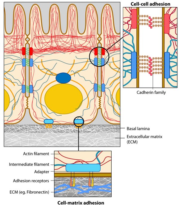
Figure: Anchoring junctions. In the illustration above, actin filaments are shown in red, microtubules are yellow, and intermediate filaments are blue. Transmembrane complexes (solid ovals) contain several different components that play a key role in linking the cell exterior to the cell interior. Cell-cell adhesion: Numerous cell-cell adhesion molecules (e.g. cadherin family of proteins) and their associated cytoplasmic anchoring components (e.g. vinculin, α-actinin) form a continuum between cells and their linked cytoskeleton components. Adherens junctions (shown as a solid red oval) primarily link actin filaments between cells, while desmosomes (shown as a solid dark-blue oval) primarily link the intermediate filaments between cells.
Cell-matrix adhesion: Interactions at CMACs and hemidesmosomes (shown as a solid light-blue oval) link the actin and intermediate filaments to the underlying matrix, respectively. Cell-matrix linkages are a key force-sensing unit that greatly influences cell polarity and migration.