
Contents
1.1 Role of Mechanobiology in Shaping Cells and Tissues 1.2 Common Themes In Mechanobiology 1.3 Types of Mechanosensing 1.4 Types of Forces Cells Encounter 1.5 The Dynamic Cytoskeleton 1.6 Common Features of Polymeric Cytoskeletal Systems 1.7 How Does the Cytoskeleton Transmit Mechanical Forces? | Essential Info: What is Mechanobiology?1.5 The Dynamic CytoskeletonThe eukaryotic cytoskeleton is a network of three long filament systems, made from the repetitive assembly and disassembly of dynamic protein components. It creates an internal architecture (see figure below) to give a cell its shape through elaborate linkage(s) to itself, the plasma membrane, and internal organelles.The cytoskeleton structure is modified by adhesion to neighboring cells or to the extracellular matrix (ECM). The strength and the type of these adhesions are pivotal for regulating the assembly/disassembly of the cytoskeleton components. This dynamic property enables cellular movement, which is governed by forces (both internal and external). This information is sensed by mechanosensors and disseminated via the cytoskeleton leading to chemical signalling and response. Components of the cytoskeletonThe primary filament systems comprising the cytoskeleton are:Although subunits of all three filament systems are present throughout the cell, differences in the subunit structures and the attractive forces between them impart each system with variable stabilities and distinct mechanical properties. These characteristics explain their distribution in particular structures and/or regions of the cell (see Figure below). Numerous cytoskeletal-associated proteins also help to regulate the spatial and temporal distribution of the cytoskeleton. The organization and assembly of one filament system is influenced by the others in a coordinated fashion for most cellular functions. Accessory proteins organize filaments into higher-order structuresCrosslinking of the filaments by specific motors or multivalent binding proteins (accessory proteins) increases stability and forms higher-order structures. Such organization facilitates generation of long-term contractile forces and occasionally support compressive forces while being dynamic. These structures are connected across cells through junctions and hence facilitate mechanotransduction and cumulative response at a tissue- or organ-level (see the lower panel in the figure below and “Mediators of mechanotransduction” for details on junctions).Accessory proteins are a critical part of the signaling network that integrates extra- and intracellular signals (e.g. force, ions etc.) with the cytoskeleton assembly module(s). These can be specific for certain types of filaments. E.g., fimbrin binds only actin filaments, while others like plectin are non-specific. Accessory factors can also help regulate the stability, mechanical properties, and force production for the individual filaments within the larger structure. For example, fascin cross links actin filaments into rigid bundles that have mechanical strength for generating protrusive force, while filamin cross links the actin filaments into gel-like networks that are flexible and produce less force. Examples of higher-order cytoskeleton structures:
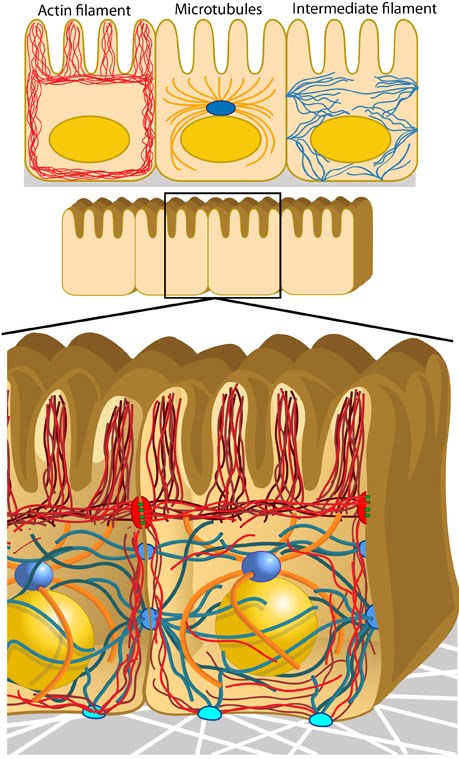
Figure: Components of the cytoskeleton. Top Panel – The cytoskeleton is composed of three main elements with distinct distribution patterns: actin filaments (red), mictrotubules (gold) and intermediate filaments (blue). Cells have
a prominent actin cortex underlying the plasma membrane that provides
mechanical strength and stability; microtubules are ordered by the
microtubule organizing center (MTOC) (navy sphere above the nucleus) that is normally just in front of
the nucleus in migrating cells and near the brush border in epithelia
(often the microtubules in epithelia are parallel with the minus ends
near the apical actin). Bottom Panel – In
epithelial cells, the location of cytoskeleton components contributes
to cell polarity, shape, and attachment of the cell to the substrate (e.g.
hemidesmosomes – light blue ovals at the bottom) or to other cells (e.g. adherens junction – red ovals on the sides, desmosome – dark blue ovals on the sides).
|
|
|
|
Current MCMF Content |
Essential Info: What is Mechanobiology? |
MBInfo © 2013 National University of Singapore.
MBInfo is sponsored by:
Sign in|Report Abuse|Print Page|Remove Access|Powered By Google Sites