Filopodium
What are Filopodia?[Edit]
Filopodia (singular filopodium) are thin membrane protrusions that act as antennae for a cell to probe the surrounding environment [1, 2, 3]. Nonprotruding filopodia are mechanistically related to microspikes [4]. Filopodia are commonly found embedded within, or protruding from the lamelliopodium at the free front of migratory tissue sheets. Filopodia are also prominent in neurite growth cones and individual cells such as fibroblasts.
 Figure 1. Different types of filopodiaFilopodia are 60-200 nm in diameter and contain parallel bundles of 10-30 actin filaments held together by actin-binding proteins (e.g. fascin). These filaments are oriented so that their barbed end is directed towards the protruding membrane.
Filopodia sense the extracellular environment at their tips using cell surface receptors [5, 6, 7]. Contact with an external target promotes the coupling of membrane-bound proteins to the backward (retrograde) flow of actin; this coupling produces the pulling forces needed for cell migration processes such as wound healing and neurite growth [8]. Contact differences between substrates or cell types influences the number of protruding filopodia [9].
A key set of proteins is involved in filopodia formation; however, the relative importance of each protein seems to vary between different organisms and their cell types. Three basic steps are involved in filopodial assembly: filament nucleation, sustained barbed end elongation and filament bundling.
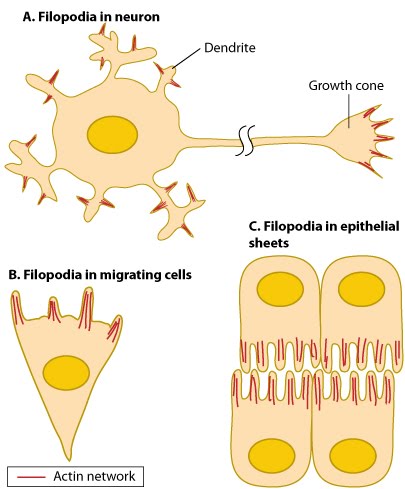 Figure 1. Different types of filopodia
Filopodia sense the extracellular environment at their tips using cell surface receptors [5, 6, 7]. Contact with an external target promotes the coupling of membrane-bound proteins to the backward (retrograde) flow of actin; this coupling produces the pulling forces needed for cell migration processes such as wound healing and neurite growth [8]. Contact differences between substrates or cell types influences the number of protruding filopodia [9].
A key set of proteins is involved in filopodia formation; however, the relative importance of each protein seems to vary between different organisms and their cell types. Three basic steps are involved in filopodial assembly: filament nucleation, sustained barbed end elongation and filament bundling.